Anti-PD-L1 antibody and application thereof

A PD-L1 and antibody technology, applied in the field of biomedicine, can solve the problems of large dosage, small range of indications, low response rate, etc., and achieve good in vivo stability and good ADCC activity

Pending Publication Date: 2021-07-16
MABWELL SHANGHAI BIOSCI CO LTD
View PDF0 Cites 2 Cited by
  • Summary
  • Abstract
  • Description
  • Claims
  • Application Information

AI Technical Summary

Problems solved by technology

[0008] As far as the currently marketed anti-PD-L1 antibody drugs are concerned, the scope of indications is small, the dosage is large, and the overall response rate is not high

Method used


Image

  • Anti-PD-L1 antibody and application thereof
  • Anti-PD-L1 antibody and application thereof
  • Anti-PD-L1 antibody and application thereof

Examples


Embodiment 1

[0107] Example 1: Preparation of anti-human PD-L1 antibody hybridoma cells

[0108] Immunization: Balb / c mice were immunized with human PD-L1 / mFc recombinant protein, and the serum titer was detected by ELISA with a 96-well microplate plate coated with human PD-L1-His recombinant protein; the serum titer reached the fusion requirement Mice were used for the next step of cell fusion.

[0109] Cell fusion and hybridoma preparation: On the 67th day after the initial immunization, select the mouse whose titer reached the requirement, aseptically take the spleen of the mouse, prepare B lymphocyte suspension, and mix it with FO myeloma cells at a ratio of 5:1. The two cells were fused under the action of PEG4000. After the fused cells were resuspended in HAT medium, they were divided into 96-well cell culture plates. Set at 37°C, 5% CO 2 Cultured in an incubator.

Embodiment 2

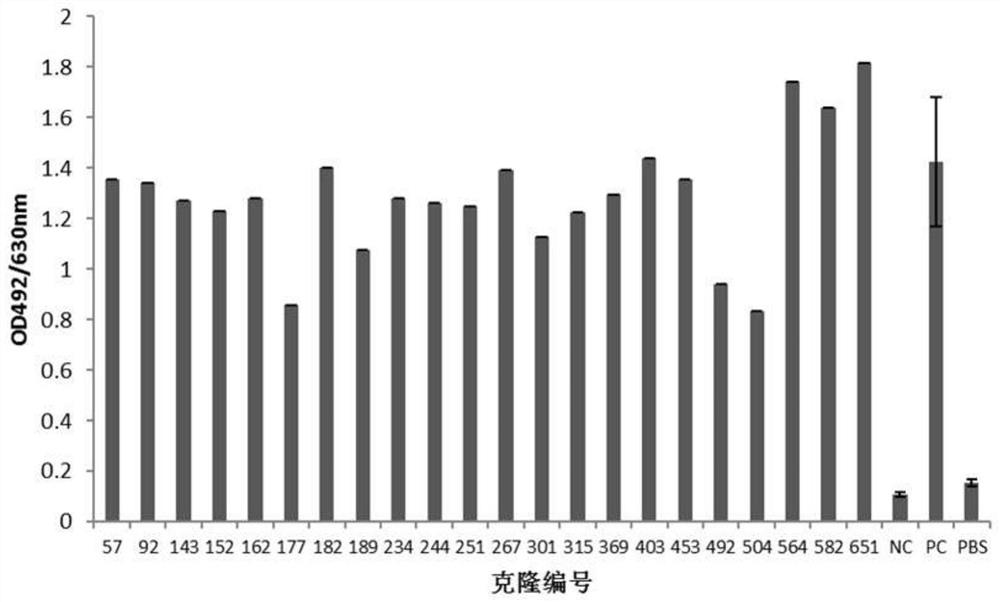
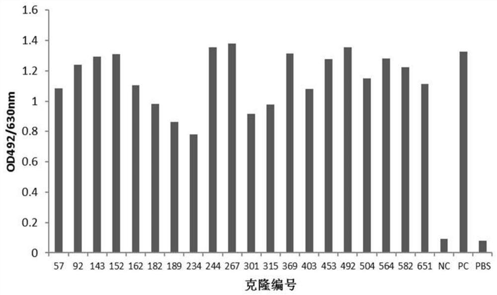
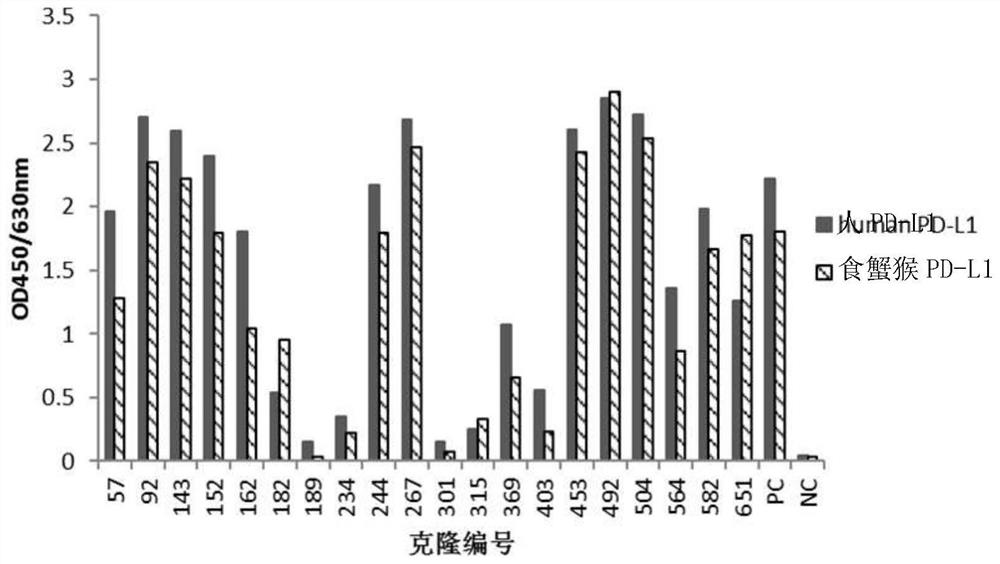
[0110] Example 2: Screening of anti-human PD-L1 antibody-positive hybridoma cell lines

[0111] 1. ELISA binding screening of positive hybridomas

[0112] 10-14 days after fusion, human PD-L1-His recombinant protein (10ug / ml, pH 9.6, 0.1M NaHCO 3 ) coated microtiter plate, 4°C, overnight; blocked with 4% skimmed milk powder-PBS, 37°C, 2h; washed three times with PBST (0.05% Tween20-PBS), added hybridoma clone culture supernatant, 37°C, 1h. Set up the following controls: (1) positive control (PC): immunized mouse serum (diluted with PBS 1:1000); (2) negative control (NC): pre-immunized mouse serum (diluted with PBS 1:1000); (3) Blank control: PBS. Washed three times with PBST (0.05% Tween20-PBS), added HRP-goat anti-mouse IgG (Fcγ), diluted 1:20000, 37°C, 1h; then washed five times with PBST (0.05% Tween20-PBS), added OPD color developing solution, avoid light for 10-15min, add 2MH 2 SO 4 Terminate the reaction; read the A492 value with a microplate reader. The A492 va...

Embodiment 3

[0122] Example 3: Sequence determination of murine anti-human PD-L1 antibody

[0123] After expanding the hybridoma cell No. 182 secreting anti-human PD-L1 antibody, use Mouse Monoclonal Antibody IgG Subclass Test Card (Cat: A12403, VicNovo) and Mouse Monoclonal Antibody Light / Heavy Chain Test Card (Cat: A12401, VicNovo) according to the reagents Subtype detection was carried out according to the operating procedures, and the subtype identification was as follows: the heavy chain was IgG1, and the light chain was Kappa chain.

[0124] Total RNA was extracted from the No. 182 hybridoma cells according to the instructions of the TRIzol kit (Cat: 15596026, Invitrogen); the total RNA of the hybridoma cells was reverse-transcribed into cDNA using M-MuLV reverse transcriptase (Cat: M0253S, NEB) ; Use degenerate primers (refer to the book [Dong Zhiwei, Wang Yan. Antibody Engineering (Second Edition). Beijing Medical University Press, 2001, 313-314]) and Phusion kit (Cat: E0553L, NE...


PUM

PropertyMeasurementUnit
Affinityaaaaaaaaaa

Abstract

The invention provides an antibody or a fragment thereof aiming at PD-L1, a coding nucleic acid of the antibody or the fragment thereof, a composition containing the antibody or the fragment thereof and the like, and application of the antibody or the fragment thereof to treatment of diseases. The antibody or the fragment thereof can specifically bind to human PD-L1 and block the binding of PD-L1 and PD-1, has a strong ADCC effect on target cells, and can significantly inhibit tumor growth.

Description

technical field [0001] The invention belongs to the field of biomedicine and relates to a novel anti-PD-L1 antibody or a functional fragment thereof. The present invention also relates to the application of the antibody or its functional fragment. Background technique [0002] Programmed death ligand 1 (PD-L1), also known as cluster of differentiation 274 (CD274) or B7 homolog 1 (B7-H1), belongs to the B7 family member and is encoded by the CD274 gene. The mature PD-L1 protein is 40kDa in size and is a type of transmembrane protein composed of 272 amino acids, which is induced and expressed in activated T cells, B cells, dendritic cells, macrophages, mesenchymal stem cells, and bone marrow-derived On the surface of mast cells and non-hematopoietic cells, and widely expressed in tumor tissues, such as lung cancer, liver cancer, bladder cancer, etc. (Dong H, Strome S E, Salomao D R et al.Tumor-associated B7-H1 promotes T-cell apoptosis: A potential mechanism of immune evasio...

Claims


Application Information

Patent Timeline
IPC IPC(8): C07K16/28C07K19/00A61K39/395A61K38/17A61K47/55A61P35/00
CPCA61K38/00A61K2039/505A61K47/55A61P35/00C07K14/71C07K16/2827C07K2317/24C07K2317/56C07K2317/565C07K2317/732C07K2317/76C07K2317/92C07K2317/94C07K2319/00A61K38/17A61K39/395C07K16/00C07K16/28C07K19/00C12N5/10C12N15/63
Inventor 王双焦莎莎王荣娟张畅
Owner MABWELL SHANGHAI BIOSCI CO LTD
Why Eureka
Try Eureka