Parasite species development stage and image pixel classification method from bottom to top

A bottom-up technology at the developmental stage, applied in image analysis, image enhancement, image data processing, etc., can solve problems such as insurmountable long-tail problems, poor detection results, and distinction of contribution weights, so as to reduce labor costs and regions Effects of medical disparities, improved parasite detection, and population suppression

Active Publication Date: 2021-08-13
BEIJING XIAOYING TECH CO LTD
View PDF7 Cites 8 Cited by
  • Summary
  • Abstract
  • Description
  • Claims
  • Application Information

AI Technical Summary

Problems solved by technology

[0011] Due to the long tail phenomenon of the parasite distribution, the loss function cannot distinguish the contribution weights of the samples of the tail category and the samples of the head category to the update of the entire model, and the simple samples and the difficult samples have the same loss function. Therefore, the biggest problem in parasite detection and classification is the long-tail problem, that is, it is easy to miss the detection of parasites, the detection effect is not good, and it is not practical

Method used


Image

  • Parasite species development stage and image pixel classification method from bottom to top
  • Parasite species development stage and image pixel classification method from bottom to top
  • Parasite species development stage and image pixel classification method from bottom to top

Examples


Embodiment 1

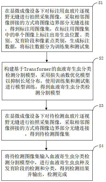
[0094] Such as figure 1 As shown, a bottom-up parasite species development stage and image pixel classification method includes the following steps:

[0095]S1. Preparation of training set and test set: under the microscope imaging equipment, the blood slides for labeling are seamlessly photographed and collected by field of view, and the border parts of the images are seamlessly connected by stitching adjacent images to obtain the image set for labeling , mark the position, category, developmental stage and pixel point category of the parasite on a single image in the image set for labeling, generate labeling data, and divide the labeling data into a training set and a test set;

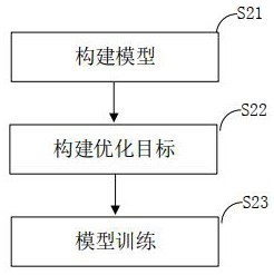
[0096] S2. Construction of Transformer-based blood parasite classification, detection and segmentation model: build a Transformer-based blood parasite classification, detection and segmentation model, use a loss function to optimize the model to suppress the long-tail distribution, use the training...

Embodiment 2

[0100] Such as figure 1 As shown, a bottom-up parasite species development stage and image pixel classification method includes the following steps:

[0101] S1. Preparation of training set and test set: under the microscope imaging equipment, the blood slides for labeling are seamlessly photographed and collected by field of view, and the border parts of the images are seamlessly connected by stitching adjacent images to obtain the image set for labeling , mark the position, category, developmental stage and pixel point category of the parasite on a single image in the image set for labeling, generate labeling data, and divide the labeling data into a training set and a test set;

[0102] S2. Construction of Transformer-based blood parasite classification, detection and segmentation model: build a Transformer-based blood parasite classification, detection and segmentation model, use a loss function to optimize the model to suppress the long-tail distribution, use the trainin...

Embodiment 3

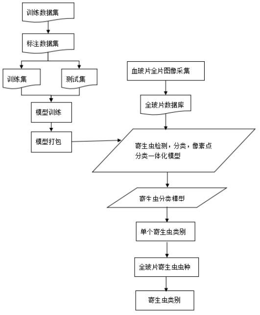
[0133] Such as image 3 As shown, a bottom-up parasite species development stage and image pixel classification method includes the following steps:

[0134] 1. Whole film collection and splicing

[0135] In order to avoid the missed detection of parasites, the present invention first seamlessly takes pictures and collects the blood slides field by field under the microscopic imaging equipment, and for the image boundary, adopts the mode of adjacent image splicing to seamlessly connect the boundary parts, so as to avoid Leave out any possible parasites.

[0136] 2. Parasite data labeling

[0137] Professional doctors use specific labeling tools to mark the position of parasites, category information and pixel category information on a single field of view. After the amount of labeling reaches a certain scale, the labeling data is divided into a training set and a test set according to a certain proportion. Learn to prepare data for network models.

[0138] 3. Construction ...


PUM


Abstract

The invention provides a bottom-up parasite species development stage and image pixel classification method, which comprises the following steps: digitalizing a slide by using a microscope or automatic slide scanning equipment, constructing a deep learning algorithm based on Transform to perform parasite detection in a view-by-view manner, and inhibiting long-tail distribution by using a focus_loss function, a CB_focal_loss function and a Jaccard function. The method is simple, parasite identification can be carried out only through the thin blood film, the detection time and labor cost can be reduced, the parasite detection level of a local area can be improved, the parasite species and the development stage can be identified, the efficiency is high, the speed is high, and the labor cost and the regional medical difference are reduced.

Description

technical field [0001] The invention relates to the technical fields of calculation, calculation and counting, in particular to a bottom-up method for classifying the developmental stages of parasite species and image pixels. Background technique [0002] Parasite detection is the main means of public prevention and control in hospitals and CDCs. Common parasites include Plasmodium, Amoeba, Leishmania donovani, Toxoplasma gondii, Babesia canis, Trypanosoma evanii, Carl's living leukocytozoa and so on. Flow cytometry screens for possible presence, not detection of parasites. [0003] At present, the detection methods of parasites mainly include: [0004] (1) Microscopic examination: Doctors use a microscope to observe peripheral blood smears to check for malaria parasites is a routine method for diagnosing malaria. Usually, it is necessary to observe the entire blood slide under a microscope with a 100x objective lens. Generally, thousands of fields of view need to be check...

Claims


Application Information

Patent Timeline
Patent Type & Authority Applications(China)
IPC IPC(8): G06T7/00G06T7/11G06K9/38G06K9/62G06N3/04G06N3/08
CPCG06T7/0012G06T7/11G06N3/08G06T2207/10056G06T2207/20081G06T2207/20084G06T2207/30004G06V10/28G06N3/045G06F18/241Y02A50/30
Inventor 李柏蕤连荷清
Owner BEIJING XIAOYING TECH CO LTD
Why Eureka
Try Eureka