Preparation method of anti-adhesion membrane for preventing and treating myophagus adhesion and anti-adhesion membrane

A technology of anti-adhesion membrane and amniotic membrane, which is applied in the field of medical materials, can solve the problems of immunogenicity, fresh amniotic membrane is not suitable for storage, transportation, and clinical difficulties, so as to reduce adhesion, promote endogenous healing of tendon, and reduce adhesion Effect

Active Publication Date: 2021-10-22
TANGSHAN WORKERS HOSPITAL
View PDF4 Cites 2 Cited by
  • Summary
  • Abstract
  • Description
  • Claims
  • Application Information

AI Technical Summary

Problems solved by technology

However, because fresh amniotic membrane is not suitable for storage and transportation, and has certain immunogenicity and other problems, it is difficult to be widely used in clinical practice.

Method used


Image

  • Preparation method of anti-adhesion membrane for preventing and treating myophagus adhesion and anti-adhesion membrane
  • Preparation method of anti-adhesion membrane for preventing and treating myophagus adhesion and anti-adhesion membrane
  • Preparation method of anti-adhesion membrane for preventing and treating myophagus adhesion and anti-adhesion membrane

Examples


Embodiment 1

[0030] A method for preparing an anti-adhesion film for preventing and treating sarcolemma,

[0031] S1. Acquisition of fresh amniotic membrane: Fresh placental tissue was bluntly separated between amniotic membrane and chorion to obtain smooth, translucent fresh amniotic membrane, soaked in balanced salt solution containing penicillin and streptomycin, and stored in a refrigerator at 4°C until use;

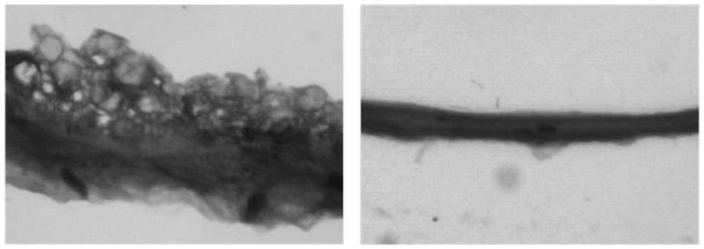
[0032] S2, preparation of decellularized amniotic membrane: fresh human amniotic membrane was rinsed with sucrose phosphate synthase (SPS) solution and cross-linked with glutaraldehyde, and the cross-linked amniotic membrane was shaken in 0.5% sodium dodecyl sulfate (SDS) 24 hours, digested with trypsin for 4 hours, rinsed thoroughly, freeze-dried, subpackaged, sterilized with ethylene oxide, cut into 1.0cm×0.5cm size for later use;

[0033] S3, loading celecoxib: accurately weigh 100mg celecoxib, grind the solid particles, completely dissolve the powder in 10mL absolute ethanol...

Embodiment 2

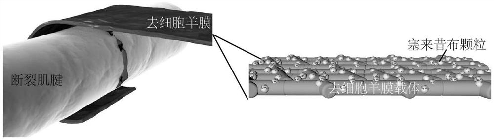
[0036] An anti-adhesion membrane for preventing and treating sarcolemma, comprising decellularized amniotic membrane on which several celecoxib particles are attached, and the size of the decellularized amniotic membrane is 1.0 cm×0.5 cm.

[0037] The decellularized amniotic membrane contains a variety of bioactive substances that can effectively promote the endogenous healing of the tendon, and celecoxib can reduce inflammation and hinder the adhesion caused by exogenous healing. The combination of the two improves the therapeutic effect of the anti-adhesion membrane and reduces the postoperative recovery time.

Embodiment 3

[0039] Drug sustained release test of celecoxib:

[0040] Accurately weigh 100 mg of celecoxib, grind the solid particles, and completely dissolve the powder in 10 mL of absolute ethanol to prepare a celecoxib mother solution with a concentration of 10 mg / mL. Then it was dissolved in PBS to prepare a series of standard solutions of celecoxib with known concentrations ranging from 0 to 0.1 mg / mL. At 249nm, the absorbance of the celecoxib standard solution with known concentration above was measured with a UV-Vis spectrophotometer (UV-2550, Shimadzu, Japan). Furthermore, the concentration (x) of celecoxib is used as the abscissa, and the absorbance (y) is used as the ordinate to map, so that the solution standard curve equation of celecoxib is: y=1.65x+0.0343, and its correlation The coefficient is r2=0.9998.

[0041] The PCL / AM nanofiber composite films with celecoxib content of 5%, 8%, and 10% were cut into sheets of 20mm×20mm, and accurately weighed. The weight was about 50...


PUM


Abstract

The invention relates to the technical field of medical materials, in particular to a preparation method of an anti-adhesion membrane for preventing and treating myophagus adhesion. The preparation method comprises the following steps: S1, acquiring a fresh amnion; S2, preparing a decellularized amnion; S3, loading the celecoxib; and S4, carrying out freeze-drying treatment. The invention also discloses an anti-adhesion membrane for preventing and treating myophagus adhesion, which is prepared by the method and comprises an acellular amniotic membrane, and a plurality of celecoxib particles are attached to the acellular amniotic membrane. The preparation method has the beneficial effects that according to the prepared anti-adhesion membrane, the amniotic membrane matrix containing various bioactive substances is loaded with celecoxib, so that endogenous healing of tendons can be effectively promoted, and adhesion caused by exogenous healing is reduced.

Description

technical field [0001] The invention relates to the technical field of medical materials, in particular to a method for preparing an anti-adhesion film for preventing and treating sarcolemma adhesion and the anti-adhesion film. Background technique [0002] With the development of industrialization, the incidence of tendon injury is increasing year by year. About 40% of patients with tendon injury will have different degrees of tendon adhesion after surgical treatment, which seriously affects the functional recovery of the affected finger, and has become one of the clinical problems to be solved urgently. [0003] The formation mechanism of adhesion is complex. During the inflammatory phase of tendon healing, a large number of inflammatory factors are released. The excessive inflammatory response mediated by it is an important reason for the formation of tendon adhesion. Therefore, many scholars use non-steroidal anti-inflammatory drugs to inhibit the adhesion-related Infla...

Claims


Application Information

Patent Timeline
Patent Type & Authority Applications(China)
IPC IPC(8): A61L31/00A61L31/14A61L31/16
CPCA61L31/005A61L31/16A61L31/146A61L2300/204A61L2300/412A61L2300/41
Inventor 刘春杰张月明白江博于昆仑田德虎
Owner TANGSHAN WORKERS HOSPITAL
Why Eureka
Try Eureka