Fixing device for living animal imaging
A technology of in vivo imaging and fixing devices, which is applied in the fields of medical science, diagnosis, diagnostic recording/measurement, etc., can solve problems such as inability to achieve long-term imaging, animals cannot continue to survive, and image instability, and achieve long-term stable imaging and weight Light, good for healing effect
- Summary
- Abstract
- Description
- Claims
- Application Information
AI Technical Summary
Problems solved by technology
Method used
Image

Examples
Embodiment 1
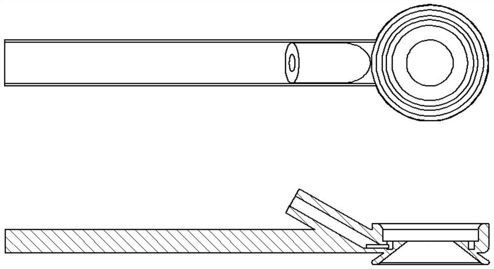
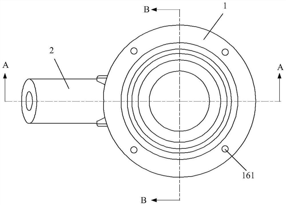
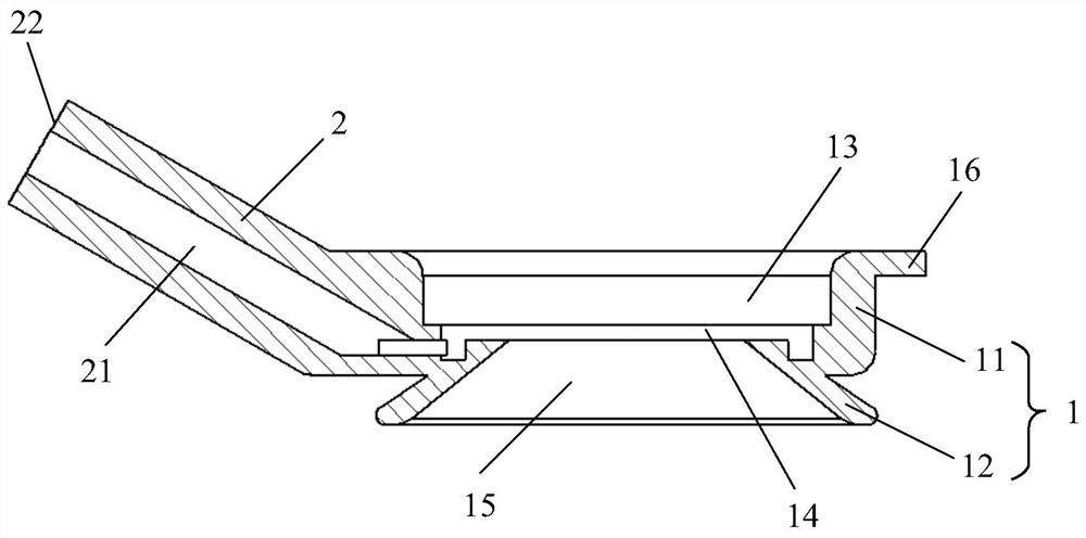
[0032] Such as Figure 2 to Figure 4 As shown, the first embodiment of the present invention provides a fixed device for live animal imaging, which includes: a hollow base 1 , an exhaust tube 2 and a cover glass. The upper part of the base 1 is a cylinder 11, and the lower part is a circular platform 12. The base 1 is provided with a cylindrical cavity 13 , a circular groove 14 and a truncated conical cavity 15 in sequence from its top surface to its bottom surface. The narrow end surface of the conical cavity 15 is in contact with the circular groove 14 . The top surface of the cylinder 11 is provided with an upper side edge 16 protruding radially from the side wall of the base 1 . A plurality of fixing holes 161 are disposed on the upper edge 16 . An air extraction pipe 2 is arranged on the side wall of the base 1, and the inside of the air extraction pipe 2 is provided with a cylindrical air extraction channel 21, which communicates with the circular groove 14 in the bas...
Embodiment 2
[0037] Such as Figure 5 to Figure 8As shown, another animal imaging fixation device provided in Embodiment 2 of the present invention. Parts in this embodiment that have the same structure as the animal live imaging and fixing device described in Embodiment 1 will not be described again. In the imaging fixing device of this embodiment, two exhaust pipes 2 distributed at 180° are arranged on the side wall of the base 1 . The number of air extraction pipes 2 and the way the air extraction pipes 2 are distributed on the side wall of the base 1 can be determined according to needs, evenly distributed or non-uniformly distributed, which is not specifically limited in the embodiment of the present invention. When using the imaging fixation device of this embodiment, the two suction tubes 6 work independently or simultaneously as required to fix the imaging tissue. The air suction port end 22 of the air extraction pipe 2 is conical, so that the hose used for air extraction is more...
Embodiment 3
[0039] Such as Figure 9 to Figure 11 As shown, there is yet another fixed device for live animal imaging provided by Embodiment 3 of the present invention. The imaging fixing device of this embodiment is provided with four exhaust pipes 2 distributed at 90° on the side wall of the base 1 of the imaging fixing device described in the first embodiment. The suction port end 22 of the suction pipe 2 is conical, and a groove 22 is arranged on the outer wall of the suction pipe 2 . During the imaging process, the four air extraction tubes 2 can work individually or in different combinations according to experimental needs to perform fixed imaging on animal tissues or organs. This structure is suitable for organs and tissues with large range of motion under physiological conditions, such as lungs and hearts. The number of air extraction pipes 2 and the way the air extraction pipes 2 are distributed on the side wall of the base 1 can be determined according to needs, evenly distrib...
PUM
 Abstract
Description
Claims
Application Information
 - R&D Engineer
- R&D Manager
- IP Professional
- Industry Leading Data Capabilities
- Powerful AI technology
- Patent DNA Extraction
Browse by: Latest US Patents, China's latest patents, Technical Efficacy Thesaurus, Application Domain, Technology Topic.
© 2024 PatSnap. All rights reserved.Legal|Privacy policy|Modern Slavery Act Transparency Statement|Sitemap