Biomarker combination for diagnosing non-obstructive azoospermia and application thereof

A biomarker, azoospermia technology, applied in the field of medical diagnosis, can solve problems such as patient pain, achieve the effect of alleviating pain, overcoming technical obstacles, and high accuracy

Pending Publication Date: 2021-12-21
温州医科大学慈溪生物医药研究院
View PDF0 Cites 0 Cited by
  • Summary
  • Abstract
  • Description
  • Claims
  • Application Information

AI Technical Summary

Problems solved by technology

[0005] At present, the method of clinical diagnosis of azoospermia is mainly through testicular biopsy, which brings great pain to the patient

Method used


Image

  • Biomarker combination for diagnosing non-obstructive azoospermia and application thereof
  • Biomarker combination for diagnosing non-obstructive azoospermia and application thereof
  • Biomarker combination for diagnosing non-obstructive azoospermia and application thereof

Examples


Embodiment 1

[0022] The construction of embodiment 1 mouse NOA model

[0023] Methods: Sexually mature male mice weighing 25-35 g were used. Dissolve busulfan in DMSO, dilute it with normal saline to obtain the dilution, disinfect the lower abdomen of the mouse with alcohol, and after drying, absorb the dilution of busulfan and inject it into the mouse intraperitoneally. After 3 injections, 35-37 days later, the mouse model of azoospermia was obtained. After sacrificing the mice, the whole blood of the mice was taken for ELISA (enzyme-linked immunosorbent assay), and the testicular tissues of the mice were taken for WB, H&E and immunofluorescence staining to test the accuracy of the mouse whole blood ELISA.

Embodiment 2E

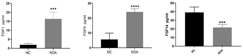
[0024] Example 2 ELISA method detects protein expression analysis of mouse NOA model blood samples

[0025] After the NOA model was established, the levels of FGF1, FGF9, and FGF14 in the serum of mice were detected by enzyme-linked immunosorbent assay kit (ELISAKit).

[0026] The operation steps of ELISA detection are as follows:

[0027] (1) Add 50 μl of FGF1, FGF9, FGF14 standard substances, serum samples and reference substances to the 96-well plate coated with the antibody, the reference substance is the blank control substance in the kit, that is, the sample diluent, Used to remove background absorbance differences caused by reagents.

[0028] The standard substance is the standard substance with different concentrations given in the kit, which is used to prepare the standard curve.

[0029] The preparation method of the serum sample is as follows: after removing eyeballs from NOA model mice, centrifuging at 3000r for 15 minutes at 4°C for 15 minutes, taking the supern...

Embodiment 3WB

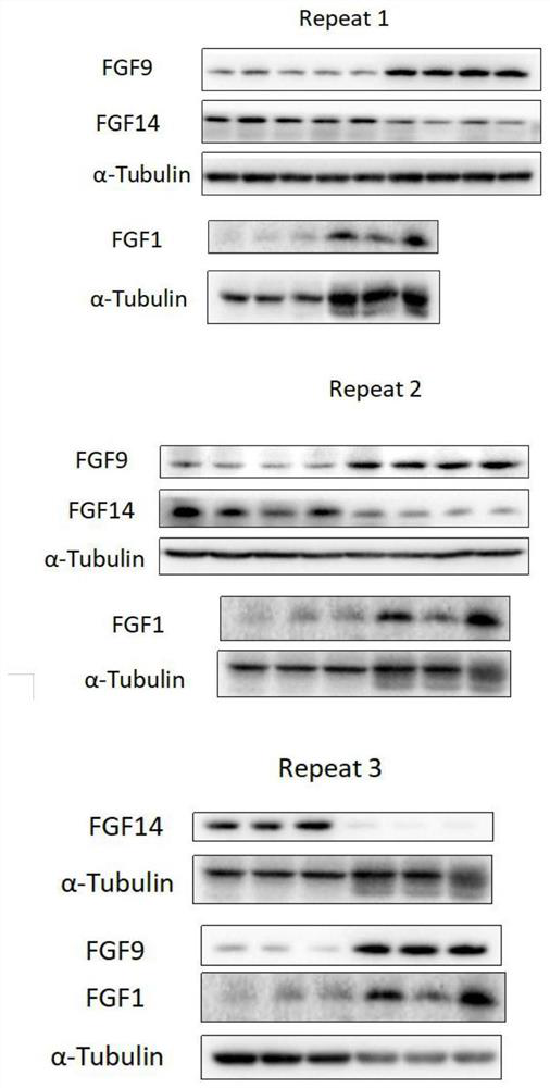
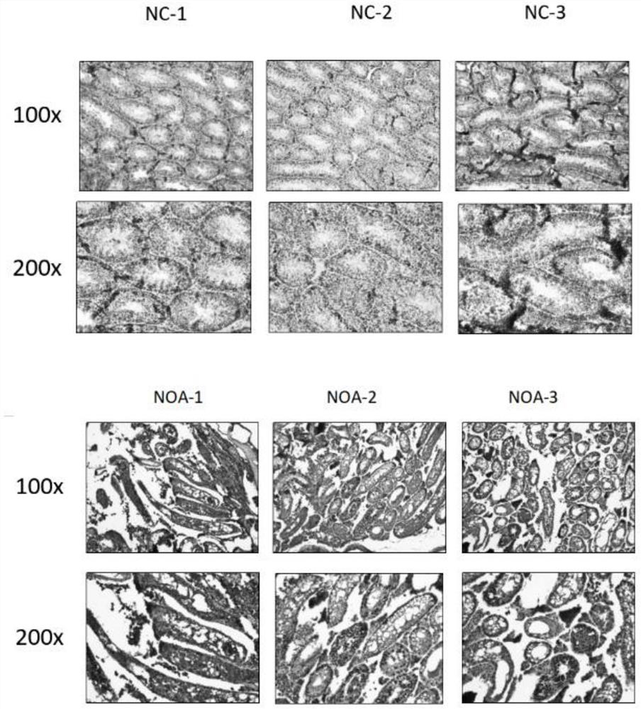
[0038] Example 3WB method, H&E method and immunofluorescence staining method detect the protein expression analysis of the mouse NOA model testis tissue sample

[0039] In order to further verify the results of protein analysis in NOA model serum, blood was collected and testicular tissues of model mice were taken for protein expression analysis by WB, H&E and immunofluorescence staining. The specific operation steps are as follows:

[0040] 1. Western blot operation steps are as follows:

[0041](1) Extraction of total protein: Take 0.05 g of NOA model mouse testis tissue, add 0.5 ml of mammalian tissue protein extraction reagent containing PMSF (extraction reagent: PMSF=100:1), and homogenize to Evenly organized. After standing on ice for 15 minutes, place it at 4°C and centrifuge at 12,000 rpm for 15 minutes, and transfer the supernatant to an EP tube.

[0042] (2) Determination of protein concentration by BCA method: Take 0, 1, 2, 4, 8, 12, 16, 20 μl of 0.5 mg / ml standa...


PUM


Abstract

The invention provides a biomarker combination for diagnosing non-obstructive azoospermia. The biomarker combination comprises FGF1 (fibroblast growth factor 1), FGF9 (fibroblast growth factor 9) and FGF14 (fibroblast growth factor 14), the biomarker combination can be used for preparing an ELISA (enzyme-linked immunosorbent assay) detection kit, the non-obstructive azoospermia is diagnosed by detecting expression conditions of FGF1, FGF9 and FGF14 in a blood sample of a patient, the pain of the patient can be greatly relieved, and the application prospect is wide.

Description

technical field [0001] The invention belongs to the field of medical diagnosis, and more specifically, the invention relates to a combination of biomarkers of non-obstructive azoospermia and its application. Background technique [0002] About 30%-55% of human infertility is related to male factors. The most common cause of male infertility is spermatogenesis disorder, which is clinically called asthenozoospermia or azoospermia. 10-15% of these are attributed to azoospermia. In recent years, with the continuous improvement of people's reproductive health needs, male infertility has increasingly become the focus of social attention. The research of the WHO Infertility Prevention and Treatment Task Force has shown that reproductive endocrine dysfunction, especially low semen quality, is an important cause of male infertility. In the past 50 years, the quality of male semen, especially the number of spermatozoa, has declined to a certain extent, and the research in our countr...

Claims


Application Information

Patent Timeline
Patent Type & Authority Applications(China)
IPC IPC(8): G01N33/68
CPCG01N33/74G01N33/6893G01N2333/50G01N2333/501G01N2800/367G01N2800/60
Inventor 龚方华张宏宇王德仲肖健李校堃
Owner 温州医科大学慈溪生物医药研究院
Why Eureka
Try Eureka