
Lateral flow analysis strip and molecular diagnostic method using same

A technology for stripping and capturing molecules, applied in chemical instruments and methods, microorganism-based methods, biochemical equipment and methods, etc. Effect

Pending Publication Date: 2021-12-24
菲尔梅迪株式会社
View PDF8 Cites 1 Cited by
  • Summary
  • Abstract
  • Description
  • Claims
  • Application Information

AI Technical Summary

Problems solved by technology

In addition, it takes a long time to perform the above-mentioned series of steps, and the diagnosis result cannot be confirmed with the naked eye
Therefore, there is a problem that it is difficult to use the method in an environment lacking a specific analysis device

Method used


Image

  • Lateral flow analysis strip and molecular diagnostic method using same
  • Lateral flow analysis strip and molecular diagnostic method using same
  • Lateral flow analysis strip and molecular diagnostic method using same

Examples


preparation example 1

[0103] Preparation Example 1: Preparation of Streptavidin-AuNP Conjugation Solution

[0104] 1 mL of 40nm AuNP (Cat.EM.GC40, BBI Solutions) solution, 100 μL borate buffer (pH8.5, 0.1M, Cat.BB001, Bio-solution) and streptavidin (Cat.434302, Thermo) was mixed to a final concentration of 10 μg / mL to prepare a mixed solution. The mixed solution was vortexed and spun down, followed by incubation at ambient temperature for 1 hour. 10 μL of PBS (10 mM, pH 7.4) containing 100 mg / mL BSA was added to the mixture solution to a final concentration of 0.1% to block the AuNP surface. The mixture was vortexed and spun down, then incubated at ambient temperature for 2 hours. The mixture was centrifuged at 9,000 rpm and 10° C. for 15 minutes using a centrifuge. The supernatant was discarded, and 1 mL of borate buffer (10 mM, pH 8.5) was added and suspended. After repeating the centrifugation and suspension process 3 times, the supernatant was removed, and 100 µL of borate buffer (10 mM) ...

Embodiment 1

[0105] Example 1: Preparation of Lateral Flow Assay (LFA) Strips

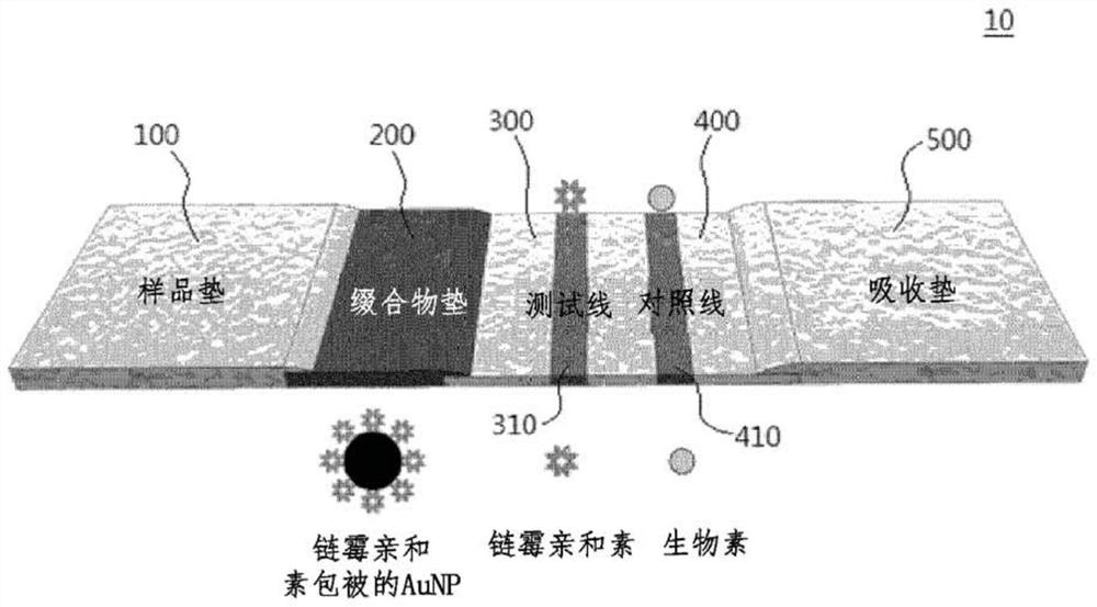
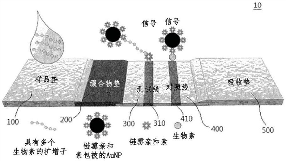
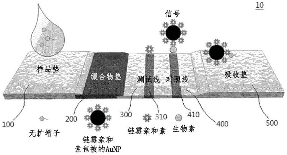
[0106] The LFA strip consists of four components: sample pad, conjugate pad, nitrocellulose membrane, and absorbent pad. Secure the components to the polyester backing card. Cut the backing card to 4 mm width using a cutting tool to prepare LFA strips. Avidin (1 mg / mL) was loaded onto the test line on the nitrocellulose membrane and biotin-BSA (1 mg / mL) was loaded onto the control line on the nitrocellulose membrane using a 1 μL pipette. The distance between these two lines is 3mm. After loading, the nitrocellulose membrane was dried at 37 °C for 1 h. 2×Avidin-AuNP conjugates were loaded onto a conjugate pad (4 mm×8 mm) and stored after drying at a temperature of 37° C. and a humidity of 25%. Thereafter, the sample pad, conjugate pad, nitrocellulose membrane, and absorbent pad were assembled in this order on an adhesive-backed card. Each section overlaps each other by 1.5mm to facilitate solution movemen...

Embodiment 2

[0107] Example 2: RT-LAMP in the presence of influenza virus RNA


PUM


Abstract

Lateral flow analysis strips and molecular diagnostic methods using the same are disclosed. The lateral flow analysis strip may comprise a sample pad (100) into which a sample comprising at least one of an amplicon labeled with a label and an amplicon precursor labeled with a label is to be introduced; a conjugate pad (200) comprising a conjugate (C1), the conjugate (C1) having an indicator and a detection agent bound to a surface of the indicator, and the conjugate (C1) being non-fixedly adsorbed to the conjugate pad (200); a test pad (300) comprising a test line (310) to which a first trapping agent is fixed; a control pad (400) comprising a control line (410) to which a second trapping agent is immobilized; and an absorbent pad (500). The lateral flow analysis strip of the present invention can detect amplicons (e.g., nucleic acids) amplified by using an amplicon precursor labeled with a type of label (e.g., a primer or dNTP) and can determine the results directly with the naked eye. Furthermore, the lateral flow analysis strip advanlabel eously has high sensitivity by using the label having a strong binding force, the trapping agent bound to the label , a detection agent, and a trapping agent bound to the detection agent. Further, by using the label having a certain binding force, the trapping agent bound to the label, the detection agent, and the trapping agent bound to the detection agent, the lateral flow analysis strip has reproducible and stable effects.

Description

technical field [0001] The present disclosure relates to lateral flow assay strips and molecular diagnostic methods using the strips. More specifically, the present disclosure relates to lateral flow assay strips capable of detecting amplicons using amplicons labeled with one type of tag and capable of diagnosis, and molecular diagnostic methods using the same. Progenitor precursors are amplified. Background technique [0002] As a conventional method for detecting and diagnosing viruses or nucleic acids, a serological method or a method using an electron microscope is mainly used. While methods using electron microscopy can confirm the presence of viruses, it is nearly impossible to identify species by their morphological characteristics. Among the serological methods, the enzyme-linked immunosorbent assay (ELISA) is the most common, but it has a detection sensitivity approximately 1,000 times lower than the polymerase chain reaction (PCR) diagnostic method and is often ...

Claims


Application Information

Patent Timeline
Patent Type & Authority Applications(China)
IPC IPC(8): G01N33/558G01N33/548C12Q1/70C12Q1/6844C12R1/93
CPCG01N33/558G01N33/548C12Q1/701C12Q1/6844C12Q2531/119C12Q2563/131C12Q2565/625C12Q1/70G01N33/54388G01N33/56911G01N33/56983G01N33/56988C12Q2527/101B01L3/5023B01L2300/069B01L2300/0825
Inventor 李柄焕金世镇金达米
Owner 菲尔梅迪株式会社
Learn More