Cell scaffold and construction method and application thereof

A cell scaffold and cell technology, applied in the field of biomedicine, can solve the problems of limited application, high cost and maintenance of 3D printing equipment, complicated operation, etc., and achieve the effects of simple operation, rapid preparation and short cycle.

Pending Publication Date: 2022-04-12
THE FIFTH AFFILIATED HOSPITAL SUN YAT SEN UNIV
View PDF1 Cites 0 Cited by
  • Summary
  • Abstract
  • Description
  • Claims
  • Application Information

AI Technical Summary

Problems solved by technology

However, the cost and maintenance of 3D printing equipment are still relatively expensive and the operation is complicated, which limits the wide application of this type of method.

Method used


Image

  • Cell scaffold and construction method and application thereof
  • Cell scaffold and construction method and application thereof
  • Cell scaffold and construction method and application thereof

Examples


Embodiment 1

[0041] In this example, cavernous mesenchymal cells were isolated.

[0042] Separation of human corpus cavernosum tissue, the corpus cavernosum tissue obtained by surgery, rinsed with PBS 3 times to remove blood, and then cut into 2mm × 2mm size block tissue, using digestive enzymes (including 4mg / mL type IV collagenase, 4mg / mL collagenase Type I collagenase, 3 mg / mL hyaluronidase and 1.5 mg / mL trypsin) were digested for 30 min, and then the mass tissue was removed with a tissue strainer, and the cell components were retained by centrifugation to obtain mesenchymal cells.

Embodiment 2

[0044] In this example, cell scaffolds were constructed and identified.

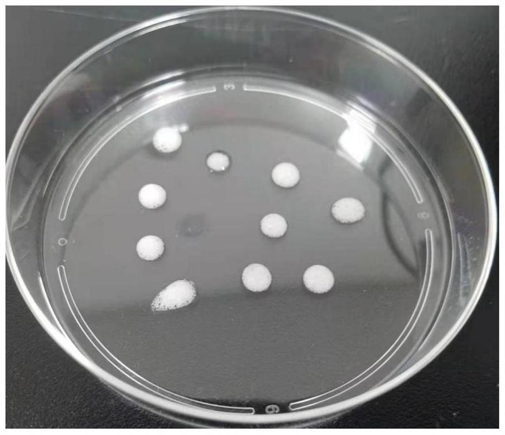
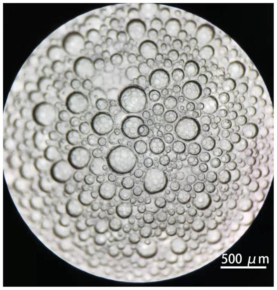
[0045] Take the mesenchymal cells isolated in Example 1, resuspend them in DMEM medium with 10% fetal bovine serum, and culture them. When the cells grow to 100% density, digest the cells to obtain a cell suspension, rinse and centrifuge with PBS, and place in Pre-cool on ice for 1 min, then use Matrigel ( 356231) resuspended at a density of 1×10 8 cells / mL, transfer 100 μL of the cell suspension to a pre-cooled 1.5mLEP tube, then immediately vortex the cell suspension, and at the same time use a 100mL pipette gun to repeatedly blow to form a large amount of foam, each time draw about 100mL of foam and place it in the on the dish (such as figure 1 Shown), under the stereoscopic microscope to observe the foam, microbubbles with a diameter of 50-500 μm can be seen ( figure 2 ), then immediately place the culture dish upside down in a 37°C incubator and wait for the matrigel to solidify. After 30 minut...

Embodiment 3

[0048] In this example, the cell scaffold prepared in Example 2 was used to culture cavernous endothelial cells.

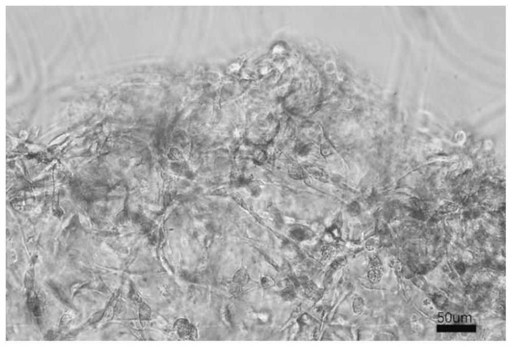
[0049] Fresh corpus cavernosum tissue was obtained surgically, digested with digestive enzymes (4mg / mL type IV collagenase, 4mg / mL type I collagenase, 3mg / mL hyaluronidase and 1.5mg / mL trypsin) for 30min, and then Use a tissue strainer to remove chunks of tissue, centrifuge to retain cell components, resuspend in endothelial cell culture medium (Lonza, EGM-2) and culture, and after 7 days, confirm the endothelial cell and smooth muscle cell colonies under the microscope according to the cell morphology, and digest them separately After forming a cell suspension, add the cell scaffold in Example 2 for co-culture, observe the colonization and growth of endothelial cells and smooth muscle in the scaffold after 2 days, mark smooth muscle cells with SAM, mark endothelial cells with CD31, and mark all nuclei with DNA staining. Separate observation and integrated observa...


PUM

PropertyMeasurementUnit
Apertureaaaaaaaaaa

Abstract

The invention discloses a cytoskeleton as well as a construction method and application thereof. The method comprises the following steps: mixing matrigel and mesenchymal cells to obtain cell gel, and culturing the cell gel to obtain the cell scaffold. The invention provides a simple, rapid and low-cost method for constructing the cell scaffold, the prepared cell scaffold has a latticed structure, the pore diameter is about 50-500 microns, the anatomical diameters of various glands and vessel tissues of a human body are covered, and the cell scaffold can be used as a growth framework of various tissue organ cells of the human body, such as kidney, prostate, micro / small blood vessels or testis, so that the cell scaffold has a good application prospect. The method is used for preparing the 3D cell culture model, and the constructed 3D cell culture model can better simulate an in-vivo physiological structure.

Description

technical field [0001] The invention belongs to the technical field of biomedicine, and relates to a cell scaffold and its construction method and application. Background technique [0002] Most of the current scientific research uses cell models and animal models. However, a large number of experimental studies have found that the above two models have certain drawbacks. On the one hand, cell models cannot simulate the complex interactions of various cells in the body. On the other hand, the species differences between animal models and humans cannot completely and intuitively reflect the changes in the human body, which limits the development of biomedicine to some extent. The emergence of 3D cell culture models has brought the possibility to make up for the above deficiencies. The 3D cell microenvironment has been proven to provide more perfect physiological conditions for cell culture, better simulate the endogenous system, and be sustainable in vitro. [0003] In recen...

Claims


Application Information

Patent Timeline
IPC IPC(8): C12N5/0775C12N5/071
Inventor 赵亮宇汤育新戴英波叶昆韩厦陈玉琢
Owner THE FIFTH AFFILIATED HOSPITAL SUN YAT SEN UNIV
Why Eureka
Try Eureka