
Novel mouse visual evoked potential recording method

A technology of visual evoked potential and recording method, applied in the medical field, can solve the problems of limited application, unusable sensor or electrode array, etc., to achieve the effect of good waveform, reliable recording method and reduced damage

Pending Publication Date: 2022-07-08
向孟清
View PDF0 Cites 0 Cited by
  • Summary
  • Abstract
  • Description
  • Claims
  • Application Information

AI Technical Summary

Problems solved by technology

However, its application is limited by the unavailability of sensors or electrode arrays in most research units

Method used


Image

  • Novel mouse visual evoked potential recording method
  • Novel mouse visual evoked potential recording method
  • Novel mouse visual evoked potential recording method

Examples


Embodiment 1

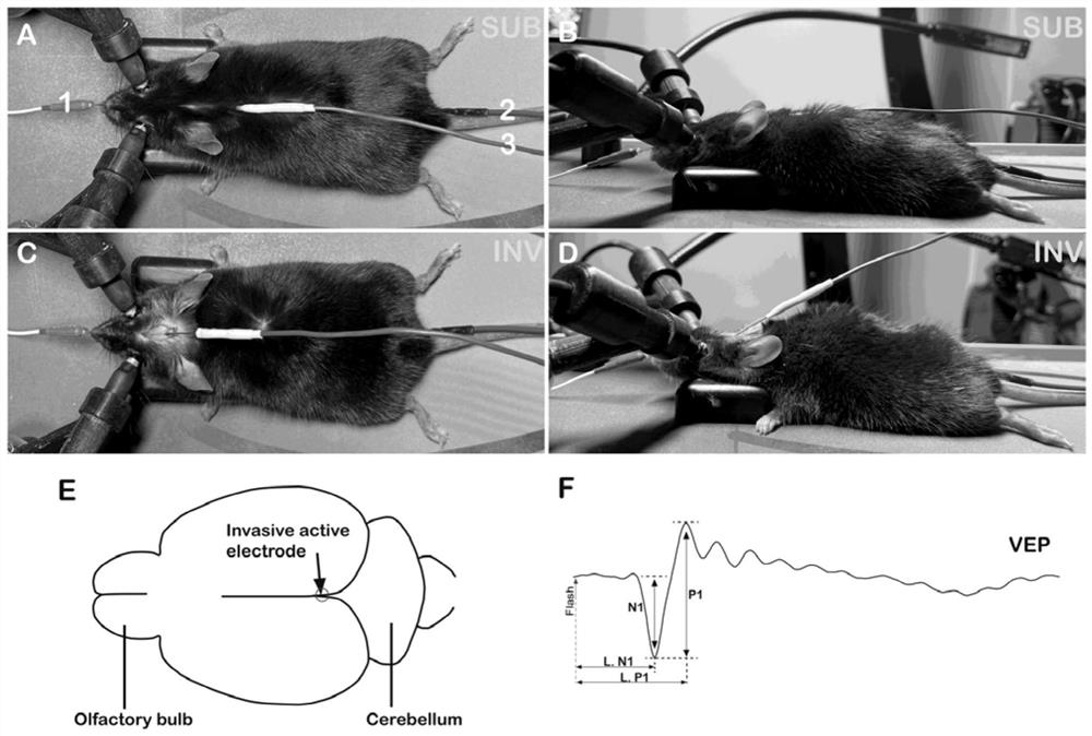
[0035] All mouse experiments were performed in accordance with IACUC (Institutional Animal Care and Use Committee) standards and approved by Sun Yat-sen University and Sun Yat-Sen Eye Center. C57BL / 6 and CD1 (CD-1, HaM / ICR) mice were purchased from Beijing Weitong Lihua Laboratory Animal Technology Co., Ltd. Brn3b AP / AP The mutant mouse is an RGC-injured mouse, and the mouse genotype was determined using standard PCR procedures. About 3 months old mice were used to record VEP. Before VEP recording, each group of mice was dark-adapted in a light-tight dark room for more than 8 hours. The test time during the day is from 8:00 to 13:00, and the test time at night is from 18:00 to 23:00. In order to keep the same dark adaptation time of different groups of mice, the dark adaptation time recorded during the day started from 20:00 the day before, and the dark adaptation time recorded at night started from 8:00 in the daytime. Mice were anesthetized with a combination of calf int...


PUM

PropertyMeasurementUnit
Impedanceaaaaaaaaaa

Abstract

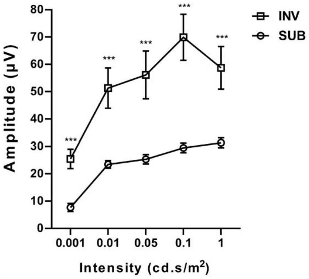
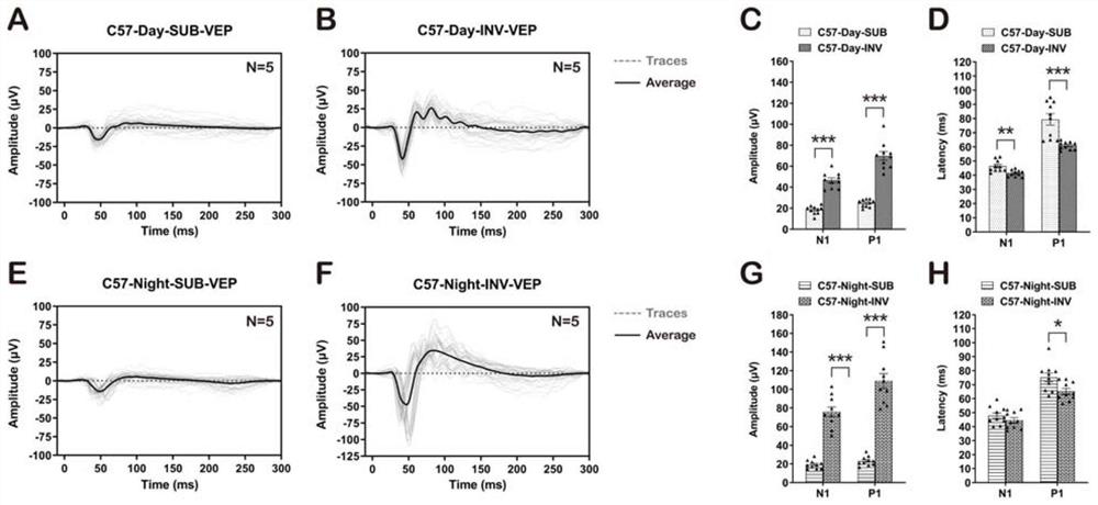
The invention discloses a novel mouse visual evoked potential recording method. According to the method, the VEP with good waveform and large amplitude can be recorded, and the influence of the position of the active electrode on the VEP is large. In a C57 or CD1 optic nerve pinch injury model mouse with retinal ganglion cell deletion and a Brn3bAP / AP mutant mouse, the VEP amplitude is obviously reduced. The method disclosed by the invention is very reliable and stable, and is particularly suitable for detecting visual response of reduced RGC quantity, dysfunction or brain connection disorder. Compared with an existing subcutaneous method (SUB), the method has the advantages that the VEP which is relatively stable, good in waveform and larger in amplitude can be recorded; compared with a skull pre-implantation screw recording method, the method has the advantage that damage of pre-implantation screws to mouse brain tissues can be reduced.

Description

technical field [0001] The invention belongs to the technical field of medicine, and in particular relates to a novel visual evoked potential recording method in mice. Background technique [0002] Visual evoked potential (VEP) is an electrical signal generated by the visual cortex in response to visual stimuli. It is an indicator of retinal function widely used in visual electrophysiology in clinical and basic research. A complete, functional visual pathway from the retina to the primary visual cortex is required (Brecelj, 2014; Ridder and Nusinowitz, 2006; Young et al., 2012). Changes in VEP parameters are closely related to physical damage as well as retinal and optic nerve pathological damage caused by genetic diseases, such as optic nerve crush (ONC), retinal detachment, diabetic retinopathy, glaucoma and other optic neuritis (Brecelj, 2014; Firan et al., 2020; Robson et al., 2018). [0003] In mice, VEP is typically recorded using hypodermic needle electrodes (Marenn...

Claims


Application Information

Patent Timeline
IPC IPC(8): A61B5/378A61B5/293
CPCA61B5/378A61B5/293A61B2503/40A61B2503/42
Inventor 向孟清刘淑婷晋康新
Owner 向孟清
Learn More