

Microfluidic channel embryo and/or oocyte handling, analysis and biological evaluation
A microfluidic, fluid channel technology, used in the determination/inspection of microorganisms, enzymology/microbiology devices, biological testing, etc., can solve problems such as the probability of inaccurate embryo damage
- Summary
- Abstract
- Description
- Claims
- Application Information
AI Technical Summary
Problems solved by technology
Method used
Image

Examples
Embodiment Construction
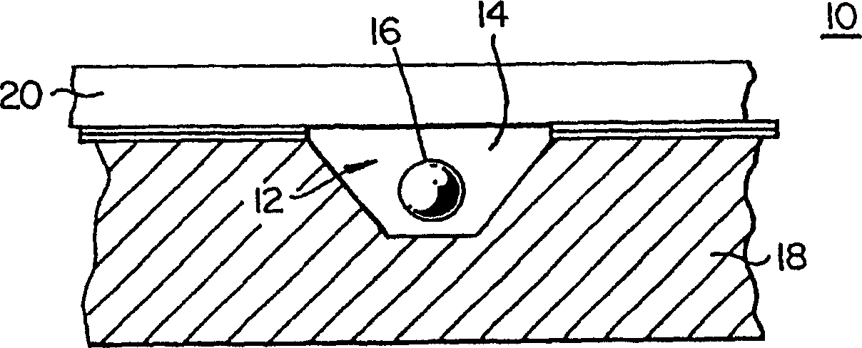
[0036] The present invention provides a microfluidic embryo handling device that reduces the stress placed on embryos when they are handled outside their natural biological host. The device and method reproduce the simulated biological rotation of an embryo through fluid-assisted movement in a channel that facilitates the sliding and rotation of the embryo. The rotation referred to here may include full rotation or partial rotation. Partial rotation may also be called shaking.
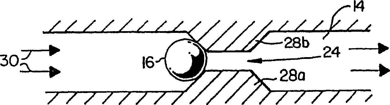
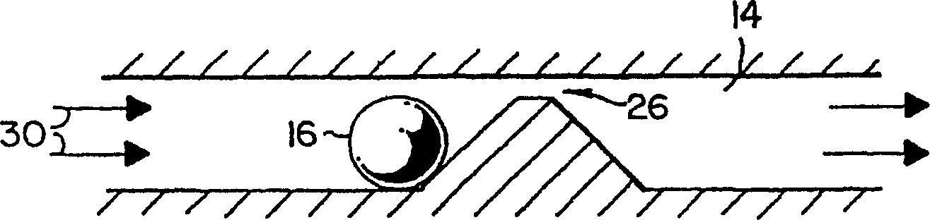
[0037] now refer to figure 1 , which is a cross-sectional view of a microfluidic embryo handling device 10 comprising an embryo transfer network 12 formed at least in part by channels 14 substantially at the embryo level. Embryos 16 within channel 14 move with the fluid flow in channel 14, while the tight dimensions of the channel cause embryos 14 to move in a simulated biological rotational motion. Channels ten times the size of embryos have been used to form rolls and slides. In biological hosts,...
PUM
| Property | Measurement | Unit |
|---|---|---|
| Diameter | aaaaa | aaaaa |
Abstract
Description
Claims
Application Information