Microflow controlled chip flow-type biochemical analysis instrument and method for detecting biochemical components

A microfluidic chip and biochemical analyzer technology, applied in the field of biochemical analyzers, can solve the problems of unstable labeled microspheres, tailing emission peaks, asymmetric peak shapes, etc., and achieves light weight, fast response speed, and reduced size. Effect of Instrument Volume

Inactive Publication Date: 2004-09-15
JILIN UNIV
View PDF0 Cites 24 Cited by
  • Summary
  • Abstract
  • Description
  • Claims
  • Application Information

AI Technical Summary

Problems solved by technology

Due to the close proximity of the excitation and emission spectra of organic dyes, and the asymmetrical peak shape, the emission peak "smears" seriously, the brightness is low and the photofading phenomenon is serious
And due to the problem of light fading, this kind of labeled microspheres is not stable and difficult to store
And it is difficult to choose the same excitation light source to excite several fluorescent dyes (generally <3 types) at the same time, so it is difficult to use them to mark a large number of microspheres. At present, there are 64 types of labeled microspheres on the market, and it is said that there will be 100 types. Microspheres with different codes are coming soon, but this is still far from enough compared to the total of about 30,000-40,000 human genes

Method used


Image

  • Microflow controlled chip flow-type biochemical analysis instrument and method for detecting biochemical components
  • Microflow controlled chip flow-type biochemical analysis instrument and method for detecting biochemical components
  • Microflow controlled chip flow-type biochemical analysis instrument and method for detecting biochemical components

Examples


Embodiment 1

[0024] Embodiment 1 further illustrates structure and working process of the present invention in conjunction with accompanying drawing

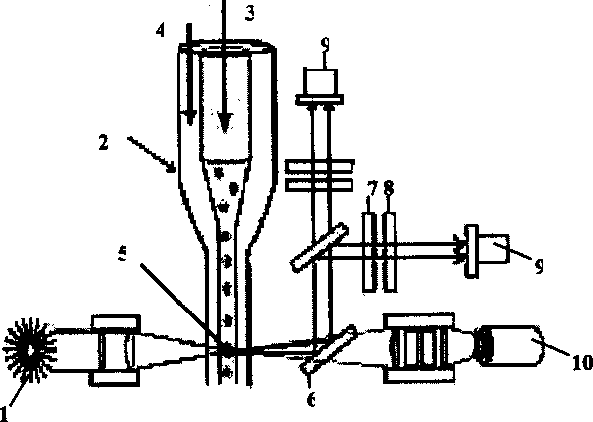
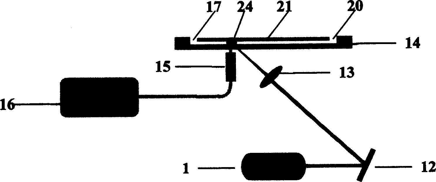
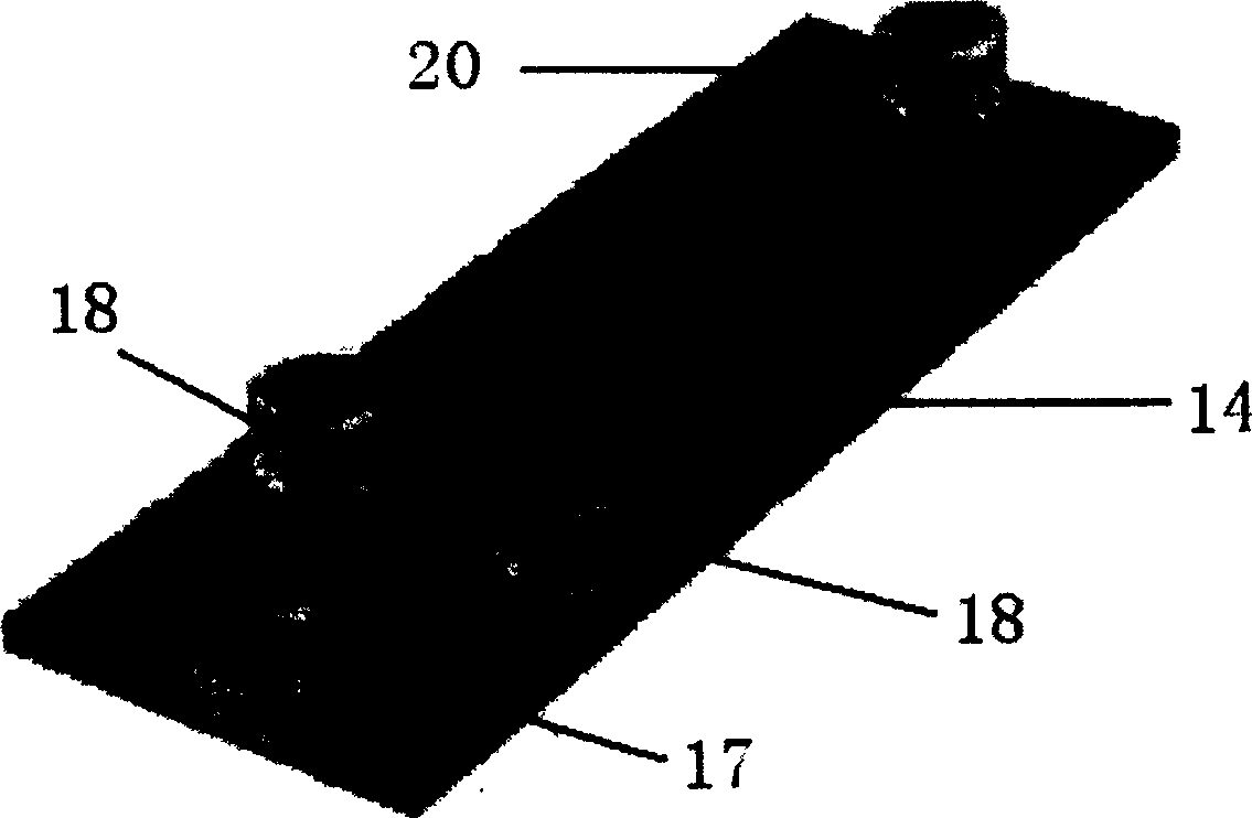
[0025]In the accompanying drawings, 1 is a laser light source, preferably a blue semiconductor laser; 12 is a plane mirror; 13 is a convex lens; 14 is a microfluidic chip, in which there are two cross channels with a width of several microns to more than one hundred microns 21. At the four end points of the microchannel 21, there are sample inflow holes 17, two opposite buffer solution inflow holes 18, and waste liquid outflow holes connected to the outside world. Sample inflow holes 17, buffer solution inflow holes 18, and waste liquid outflow Corresponding liquid reservoirs are respectively bonded on the holes 20, and an electrode is inserted in each of them; 24 is a detection area, which is located on the channel 21 leading to the waste liquid outflow hole 20; The detection point 24 is opposite, and the other end is closely connected with...

Embodiment 2

[0033] Example 2 Detecting the antigen contained in the sample:

[0034] 1. Shake the coded microspheres in a vortex shaker and an ultrasonic shaker to suspend them evenly; 3-(3-Dimethylaminopropyl) carbodiimide salt (EDC) was mixed and shaken, and left at room temperature for 20 minutes to activate the carboxyl groups on the surface of the microspheres.

[0035] 2. The activated microspheres were washed 3 times with PBS solution, then suspended by vortex, and the capture antibody was added quickly, after mixing, shake and incubate at room temperature for 30min, centrifuge at 200g for 15min, and discard the supernatant.

[0036] 3. Suspend the microspheres in BSA containing 1mg / mL and 0.02% Tween 20, incubate at 4°C for 30min, centrifuge at 200g for 15min, discard the supernatant; wash the microspheres twice with the above buffer, and suspend them in Incubate in 1 mL of cell culture medium at 4°C for 20 min; count with a hemocytometer, and adjust the concentration of suspende...

Embodiment 3

[0040] Example 3 Detection of cytokines by microsphere-labeled receptors

[0041] 1. The process of activating the hydroxyl groups on the surface of the microspheres is the same as in Example 2.

[0042] 2. Add receptors such as IL-1, 2, 6, and 12 to each microsphere, each microsphere corresponds to a receptor, mix and incubate at room temperature for 30 minutes, centrifuge at 200g for 15 minutes, and discard the supernatant ;

[0043] 3. Suspend the microspheres in BSA containing 1mg / mL and 0.02% Tween 20, incubate at 4°C for 30min, centrifuge at 200g for 15min, discard the supernatant; wash the microspheres twice with the above buffer, and suspend them in Incubate in 1 mL of cell culture medium at 4°C for 20 min; count with a hemocytometer, and adjust the concentration of suspended microspheres to 2×10 6 / mL, and placed in a dark place at 4°C;

[0044] 4. Add 100 μL of the solution containing the cytokine to be tested, incubate at 4°C for 45 minutes; centrifuge at 200 g f...


PUM


Abstract

Microflow controlled chip 14 is prepared by sticking etched two layers of sheets. Chunnel 21 in decussation is fluted between the said sheets. There are inhalant pore 17 of sample, opposite two inhalant pores 18 for buffer solution, and flowout pore for waste fluid at end point of channel 21 connected to outside. One electrode is inserted into the four pores respectively. Laser emitted from laser source 1 is focused on testing point 24 on the channel 21 in decussation. Fluorescence emitted from matter in channel 21 and received by optical fiber is transferred to spectrum detection system 16 composed of CCD spectrographic detector. The invention possesses features of small size, lightweight, compact structure, and simple operation, as well as optional fluorescent reagent, fast response speed and fluorescence intensities in multiple wavelengths obtained. The invention is applicable to areas such as analyzing biochemistry matter, drug screening and clinical diagnosis.

Description

technical field [0001] The invention belongs to a biochemical analyzer used in biochemistry, pharmaceutical research, biomedical research and clinical diagnosis, etc. and a method for detecting nucleic acid, antibody or antigen, peptides and chemical factors using the biochemical analyzer. Background technique [0002] The prior art close to the present invention is organic fluorescent dye-encoded microspheres and flow cytometry based on fluorescent dye-encoded microspheres. Quantitative flow cytometry based on fluorescent dye-labeled microspheres has been used to accurately quantify cell surface receptors, etc., and the results are comparable to traditional Scatchard assays and can be used for inter-laboratory standardized studies, As a result, several companies have started to produce high-quality fluorescent calibration beads. [0003] Instruments that combine organic fluorescent dye-encoded microspheres with traditional flow cytometers for simultaneous detection of mult...

Claims


Application Information

Patent Timeline
IPC IPC(8): C12Q1/68G01N21/64G01N27/26G01N33/50G01N33/68G01N35/08
Inventor 牟颖金钦汉杨蕊吴喆林章碧
Owner JILIN UNIV
Why Eureka
Try Eureka