

Method for separating silk gland cell of silkworm suited to researches in molecular biology
A molecular biology, silkworm technology, applied in biochemical equipment and methods, animal cells, microorganisms, etc.
- Summary
- Abstract
- Description
- Claims
- Application Information
AI Technical Summary
Problems solved by technology
Method used
Image

Examples
Embodiment 1
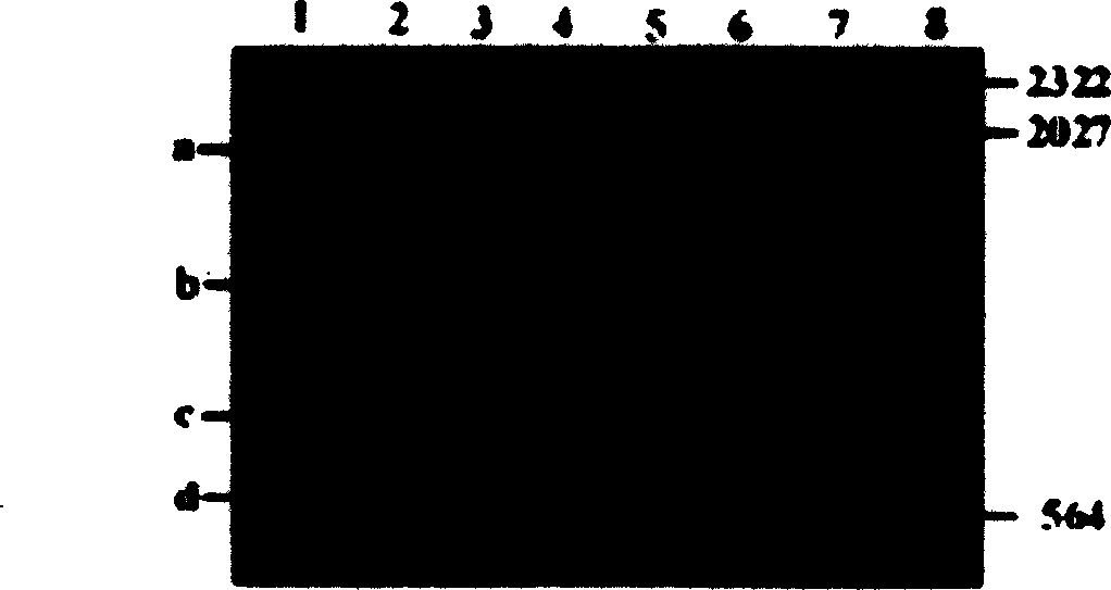
[0019] Example 1: PCR reaction results of posterior silk gland cells
[0020] (1) In a 0.7% NaCl solution at 4°C, dissect the rear silk glands of the five-instar, fifth-day silkworm of the Qiufeng variety frozen in liquid nitrogen, wash thoroughly with 0.7% NaCl solution, and transfer to pre-cooling After dipping in 60% ethanol for 5 minutes, fix one end of the silk gland with a pair of tweezers, and use another pair of tweezers to gently hold the surface of the silk gland while gently pulling to peel off the posterior silk gland cells.
[0021] (2) After drying the posterior silk gland cells in a vacuum desiccator, add 20 μl 0.3mol / L NaOH, 1% ME buffer solution to each 1mg posterior silk gland cells, grind in ice bath for 10min, add 50μl 60mmol / LTris-HCl pH 8 Buffer solution, stirred for 10 minutes, centrifuged at 15000×g, 4°C for 5 minutes. Take 20 μl of the supernatant, add 50 μl of 99% ethanol, place at -20°C for 15 minutes, centrifuge at 15,000×g at 4°C for 15 minutes, d...
Embodiment 2
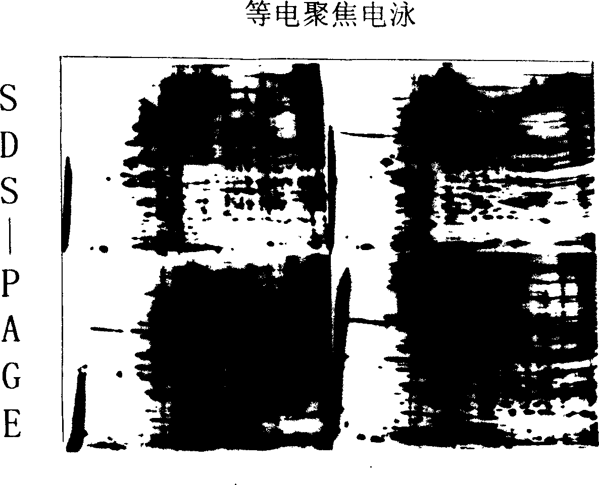
[0026] Example 2: Results of Two-dimensional Electrophoresis of Proteins in Central Silk Gland Cells
[0027] (1) In 0.7% NaCl solution at 4°C, dissect silkworms of five instars and different days of P50 variety frozen in liquid nitrogen to obtain the middle silk glands of silkworms, wash them thoroughly with 0.7% NaCl solution, and transfer them to pre-cooling After dipping in 60% ethanol for 5 minutes, fix one end of the silk gland with a pair of tweezers, and gently pull the surface layer of the silk gland with another pair of tweezers to peel off the silk gland cells in the middle.
[0028] (2) Put the central silk gland cells into a weighed centrifuge tube, dry them in a vacuum desiccator, and weigh the central silk gland cells. Add 10 times (μl) of Tris-HCl extraction buffer (pH 9.5, 40mM) to the weight of silk gland cells (mg) in portions, grind thoroughly on ice, sonicate for 2 minutes, and place in an ice bath for 10 minutes.
[0029] (3) Centrifuge twice at 4° C. at...
Embodiment 3
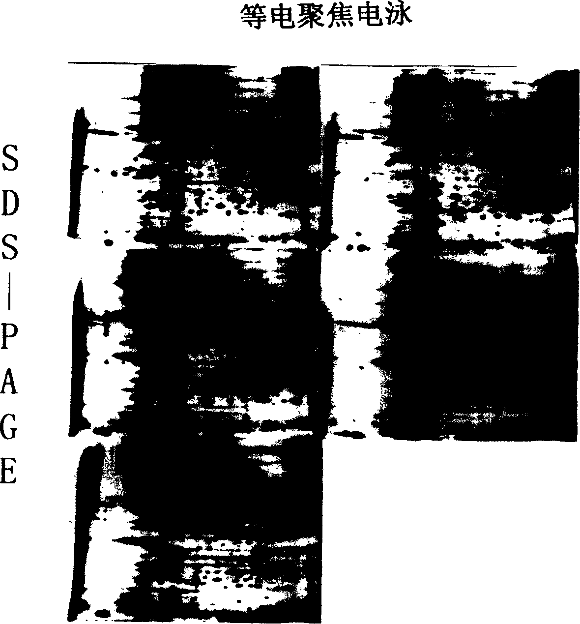
[0035] Example 3: Results of Two-dimensional Electrophoresis of Proteins in Central Silk Gland Cells
[0036] (1) In 2% NaCl solution at 20°C, dissect fresh silkworms of five instars and different days of the P50 variety to obtain silk glands in the middle part of the silkworm, fully wash them with 0.8% NaCl solution, and then transfer them to pre-cooled 80% silkworm After immersing in ethanol for 2 minutes, fix one end of the silk gland with a pair of tweezers, and use another pair of tweezers to gently hold the surface layer of the silk gland while gently pulling to peel off the silk gland cells in the middle.
[0037] (2) Put the central silk gland cells into a weighed centrifuge tube, dry them in a vacuum desiccator, and weigh the central silk gland cells. Add 10 times (μl) of phosphate extraction buffer (PBS) (32.5mM K 2 HPO 4 , 2.6mM KH 2 PO 4 , 400mM NaCl, pH 7.6), thoroughly ground on ice, ultrasonicated for 2 minutes, and placed in an ice bath for 10 minutes.
[...
PUM
 Abstract
Description
Claims
Application Information