Cell imaging method based on laser induced fluorescence

A laser-induced fluorescence and cell imaging technology, which is applied in fluorescence/phosphorescence, material excitation analysis, etc., can solve the problems of weak signal detection difficulties, insufficient concentration of excitation light source energy, and non-single spectral lines of mercury lamps, etc.

Inactive Publication Date: 2005-12-28
DALIAN INST OF CHEM PHYSICS CHINESE ACAD OF SCI
View PDF0 Cites 9 Cited by
  • Summary
  • Abstract
  • Description
  • Claims
  • Application Information

AI Technical Summary

Problems solved by technology

Using a fluorescence microscope as the core equipment is a very common and commonly used method for cell fluorescence imaging. However, due to factors such as the non-single spectral line of the mercury lamp and the insufficient concentration of the excitation light source, it is difficult to detect weak signals.

Method used


Image

  • Cell imaging method based on laser induced fluorescence
  • Cell imaging method based on laser induced fluorescence
  • Cell imaging method based on laser induced fluorescence

Examples


Embodiment 1

[0024] Example 1 (Dye Influx Cell Assay)

[0025] The subcultured suspension cells were transferred to centrifuge tubes, washed twice with phosphate buffer, and then the centrifuged cells were diluted with phosphate buffer supplemented with Rho123 (13.1 pM). Incubate the cell solution in a 37°C incubator for a certain period of time. After centrifugation, wash twice with phosphate buffer containing transporter inhibitors. Make the cell concentration at 10 5 Amounts / ml or so, as a sample for fluorescence imaging.

[0026] With reference to the position of the cell in the optical photo, the luminescence intensity values ​​of 10 pixels are taken from each cell in the fluorescence photo, and the sum is used as the luminescence intensity of the cell at this position. Take more than 10 cells in each fluorescence photo to calculate the average luminescence intensity.

[0027] Sensitive cells (S, cell line with low rhodamine transporter expression) and drug-resistant cells (A, cel...

Embodiment 2

[0028] Example 2 (dynamic observation of the process of sugar molecules entering plant cells)

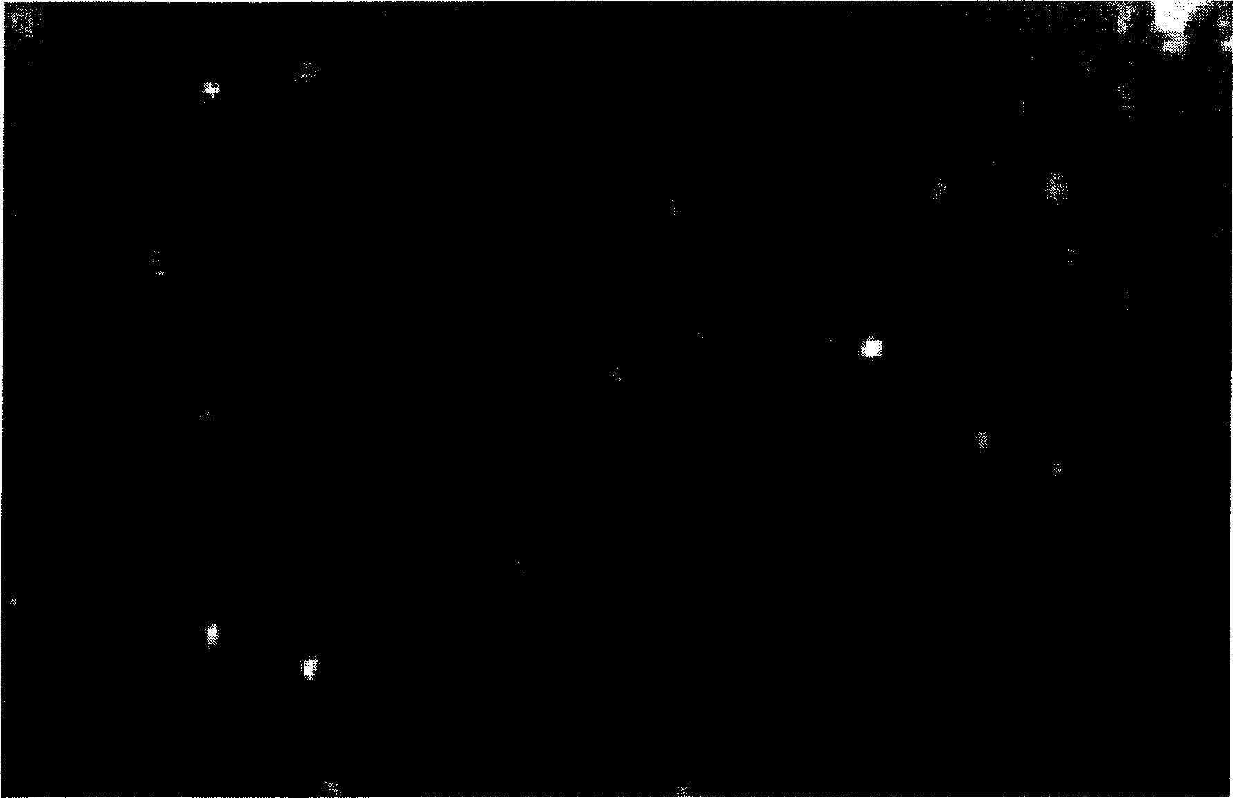
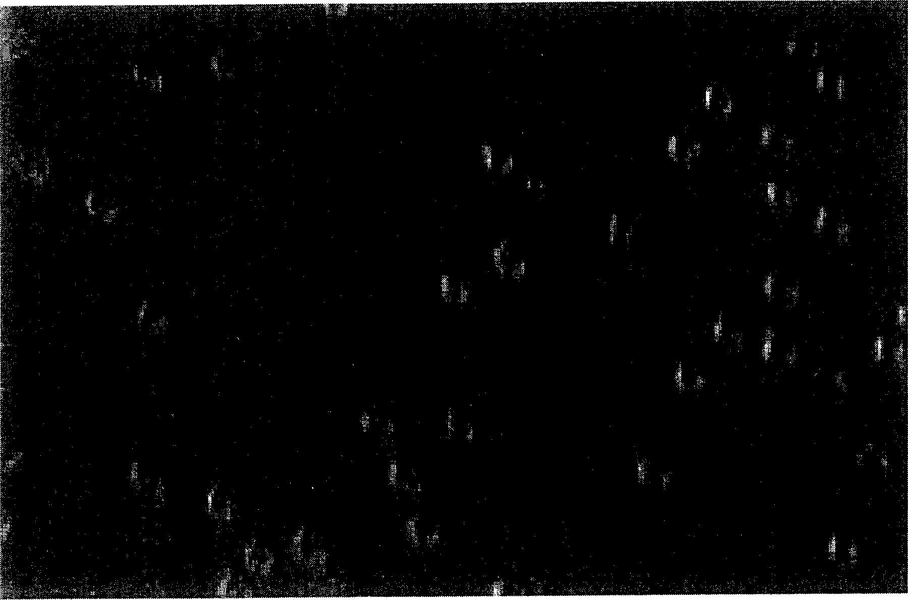
[0029] Take 10uL of cultured plant cell fluid (the cell concentration is 10 5 1 / ml or so, as a sample substrate for fluorescence imaging), drop it on the prism, add 1uL of pre-labeled oligosaccharides with dyes, open the shutter only during the exposure time of the monitor, and record a frame of cell fluorescence photos every 10 seconds ( See Figure 4 ).

[0030] Figure 4 The middle photos 1 and 2 are the first 20 seconds of photos, and the plant cells are relatively blurred. After 30 seconds, the local fluorescence intensity of individual cells increased. With further increase in time, the entire cell fluoresces (see Figure 4 in photo 9). This process shows the dynamics of the entry of sugar molecules into plant cells.


PUM


Abstract

A method of cell imaging based on using laser to induce fluorescent light collects fluorescent light emitted from dye - labeled cell by objective lens of microscope under shine of laser to be cell be image in CCD monitor through filter and then to obtain photograph of the cell. The light path portion of device for realizing the method consists of laser, shutter, extender lens, reflector, prism, objective lens of microscope, trap filter, band - pass filter and CCD monitor.

Description

Technical field: [0001] The invention mainly relates to a cell imaging method, and particularly provides a cell imaging method based on laser-induced fluorescence. Background technique: [0002] Fluorescence imaging of cells plays an important role in observing the morphological structure of living cells, monitoring the interaction between cells and external molecules, and quantifying the content of intracellular substances. [0003] The existing cytofluorescence imaging methods mainly fall into two categories: [0004] A confocal scanning microscope and a total internal reflection microscope with laser as the excitation light source are used for operation. They usually operate directly by using the laser light emitted by the laser as the excitation light source. As a mature technology, cell imaging operations with laser confocal scanning microscope as the core equipment have been widely used in biology, medicine and other fields. In this technique, a laser spot focused t...

Claims


Application Information

Patent Timeline
Patent Type & Authority Applications(China)
IPC IPC(8): G01N21/64
Inventor 盖宏伟王玉林赵小明马银法林炳承
Owner DALIAN INST OF CHEM PHYSICS CHINESE ACAD OF SCI
Why Eureka
Try Eureka