

Preparation method of tissue engineering bone cartilage compound and its application
A tissue-engineered bone and composite technology, used in biochemical equipment and methods, bone implants, tissue cell/virus culture devices, etc., to achieve the effects of promoting early formation, delaying degeneration, and cartilage stabilization
- Summary
- Abstract
- Description
- Claims
- Application Information
AI Technical Summary
Problems solved by technology
Method used
Image

Examples
preparation example Construction
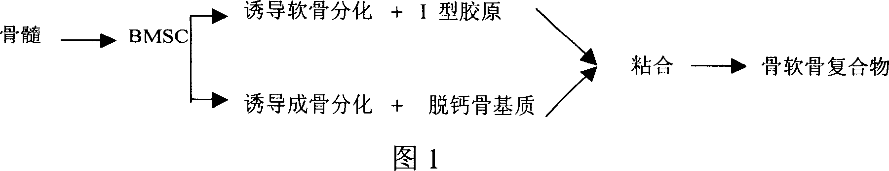
[0038] As shown in Figure 1, the tissue engineering osteochondral compound preparation method of the present invention comprises the following steps:
[0039]1) Use the method of density gradient centrifugation of lymphocyte separation medium to isolate and expand bone marrow pluripotent stem cells BMSCs in vitro. When BMSCs reach confluence at passage 2-3, digest and count according to 3×10 7 -6×10 7 / 100mm 2 Density inoculation, using different cartilage and osteogenesis induction systems for initial induction of cartilage and bone origin;
[0040] 2) taking fresh ilium and knee joints of New Zealand rabbits, and performing decalcification treatment to prepare decalcified bone matrix;
[0041] 3) Then the demineralized bone matrix and type I collagen sponge were cut into cubes of required size, pre-coated with human fibronectin and dried overnight;
[0042] 4) BMSCs cultured in different induction systems in step 1 were respectively inoculated into the decalcified bone ma...
Embodiment 1
[0046] Example 1 Using mesenchymal stem cells to prepare tissue-engineered cartilage to repair articular cartilage defects
[0047] 1) In vitro isolation and expansion of bone marrow pluripotent stem cells: extract rabbit bone marrow through bone marrow puncture, and use density gradient centrifugation to separate mononuclear cells with lymphocyte separation medium (specific gravity: 1.077) 7 / 100mm 2 The density was inoculated in culture flasks, and the culture medium contained 60% DMEM-LG / F / 12, 40% MCDB-201 (Gibco), 2% FBS, 10ng / ml EGF, 10ng / ml PDGF-bb (Peprotech) components . Place at 37°C, 5% CO 2 cultured in an incubator. After 24 to 72 hours, discard the suspended cells, add fresh medium to continue culturing, and change the medium every 3 days until the cells grow to 80% confluence, digest the cells with 0.25% trypsin-0.1% EDTA at 37°C, Count to 3 x 10 4 -5×10 4 / cm 2 Cells were inoculated at a certain density, and cultured and subcultured according to the above ...
Embodiment 2
[0052] Example 2 Using Bone Marrow Mesenchymal Stem Cells to Prepare Tissue-Engineered Osteochondral Compounds to Repair Articular Cartilage Defects
[0053] 1) In vitro isolation and expansion of bone marrow pluripotent stem cells: the rabbit bone marrow was extracted by bone marrow puncture, and the bone marrow mononuclear cells were separated by lymphocyte separation medium. 7 / 100mm 2 The density was inoculated in culture flasks, and the culture medium contained 60% DMEM-LG / F / 12, 40% MCDB-201 (Gibco), 2% FBS, 10ng / ml EGF, 10ng / ml PDGF-bb (Peprotech) components . Place at 37°C, 5% CO 2 cultured in an incubator. After 2-3 days, discard the suspended cells, add fresh medium to continue culturing, and change the medium every 3 days until the cells grow to 80% confluence, digest the cells with 0.25% trypsin-0.1% EDTA at 37°C, and count , to 3 x 10 4 -5×10 4 / cm 2 Cells were inoculated at a certain density, and cultured and subcultured according to the above conditions. ...
PUM
 Abstract
Description
Claims
Application Information