Medical adhesive and methods of tissue adhesion

A technology of adhesives and organic tissues, applied in the direction of surgical adhesives, applications, medical science, etc., to achieve the effects of easy synthesis, reduced possibility, and strong tensile strength

Inactive Publication Date: 2007-05-23
UNIVERSITY OF PITTSBURGH
View PDF0 Cites 14 Cited by
  • Summary
  • Abstract
  • Description
  • Claims
  • Application Information

AI Technical Summary

Problems solved by technology

Assuming that the penetration depth of light is limited, radiation curing limits the application of this technique to films that are susceptible to light sources

Method used


Image

  • Medical adhesive and methods of tissue adhesion
  • Medical adhesive and methods of tissue adhesion
  • Medical adhesive and methods of tissue adhesion

Examples


Embodiment 1

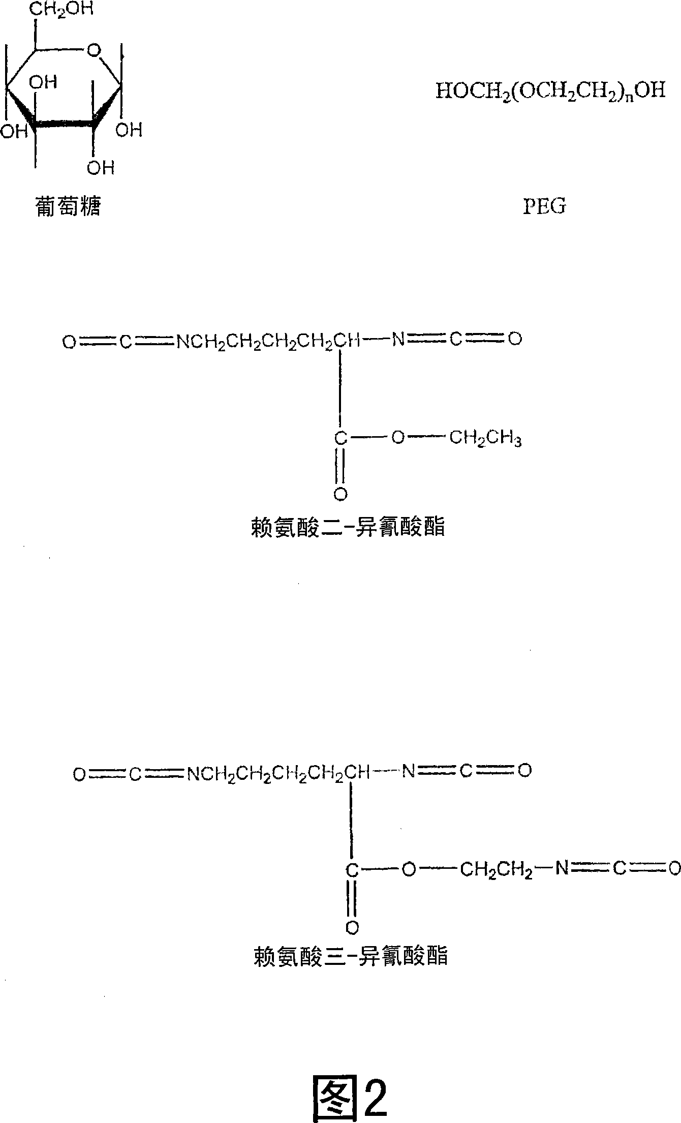
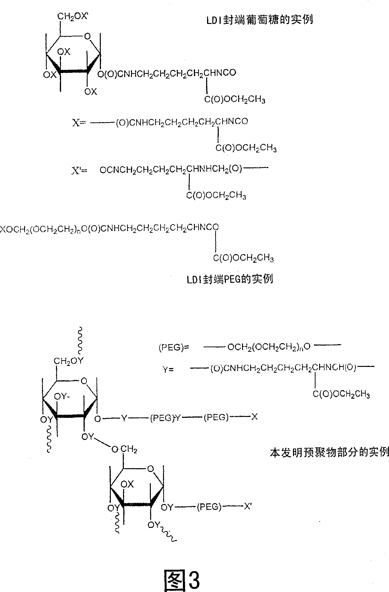
[0045] Representative LDI-based polyurethane tissue adhesives or stickies were synthesized using the procedure described below. To prepare the adhesive, 0.5889 g of glucose (3.27 mmol, -OH 16.36 mmol) was added to 5 ml of PEG 400 (14.09 mmol, -OH 28.18 mmol) in a dry round bottom flask, purged with nitrogen and heated at 50 Heated at ℃ to obtain a transparent solution. PEG is liquid at room temperature and dissolves glucose without the need for additional solvents. Then, 4.6 ml of lysine di-isocyanate (LDI, d 1.157, FW 226, 23.55 mmol, -NCO 47.10 mmol) were added, and the flask was fitted with a rubber septum and sealed. The reaction mixture was stirred at 50 °C for 48 hrs, resulting in a viscous solution. Store the viscous solution under nitrogen at room temperature until use. The viscous liquid is spread on each of the two wet tissues, and when the two tissues are pressed together, they adhere firmly to each other after about 1-2 minutes.

Embodiment 2

[0047] Using PEG200 instead of PEG400, another LDI-based polyurethane tissue adhesive was synthesized using the procedure described below, which ultimately resulted in a harder, stronger seal than the adhesive of Example 1. During this process, 0.6 g of glucose (3 mmol, -OH 15 mmol) was added to 5 ml of PEG 200 (28.18 mmol, -OH 56.35 mmol) in a dry round bottom flask, purged with nitrogen and heated at 50 °C, A clear solution was obtained. Then, 7ml of LDI (d 1.157, FW 226, 35.83mmol, -NCO 71.67mmol) was added, and the flask was fitted with a rubber septum and sealed. The reaction mixture was stirred at 50 °C for 48 hrs, resulting in a viscous solution. Store stickies under nitrogen at room temperature until use. Spread the viscous liquid over each of the two wet tissues, and when the two tissues are pressed together, they adhere firmly to each other after 1-2 minutes.

Embodiment 3

[0049] Example 3 shows that when the glucose fraction in the reaction mixture is increased, the time required to close the wound is shorter, the bond strength is increased and the final material is stiffer. In this study, 1.8 g of glucose (10 mmol, -OH 50 mmol) was added to 5 ml of PEG 200 (28.18 mmol, -OH 56.35 mmol) in a dry round bottom flask, purged with nitrogen and heated at 50 °C, A clear solution was obtained. Then, 10 ml of LDI (d 1.157, FW 226, 51.19 mmol, -NCO 102.02 mmol) were added. The flask was fitted with a rubber septum and sealed. The reaction mixture was stirred at 50 °C for 48 hrs, resulting in a viscous solution. The stickies were stored under nitrogen at room temperature until use. The viscous liquid was allowed to spread over each of the two wet tissues, and when the two tissues were pressed together, they adhered firmly to each other after about 1 minute.


PUM

PropertyMeasurementUnit
Functional group degreeaaaaaaaaaa

Abstract

An adhesive including a mixture of isocyanate capped molecules formed by reacting multi-isocyanate functional molecules with multi-functional precursor molecules including terminal functional groups selected from the group consisting of a hydroxyl group, a primary amino group and a secondary amino group. Preferably, the functional groups are hydroxyl groups. The multi-functional precursor compounds are biocompatible. Multiamine functional precursors of the multi-isocyanate functional molecules are also biocompatible. As discussed, above, the mixture of molecules preferably has an average isocyanate functionality of at least 2.1 and, more preferably, has an average isocyanate functionality of at least 2.5. As also described above, the mixture of molecules preferably has a viscosity in the range of approximately 1 to approximately 100 centipoise. The mixture of molecules forms a crosslinked polymer network upon contact with the organic tissue in the presence of water. The crosslinked polymer network is biocompatible and biodegradable. The crosslinked polymer network degrades into degradation products including the precursor molecules and the multi-amine functional precursors.

Description

technical field [0001] The present invention relates generally to medical adhesives and methods of tissue closure, and more particularly to medical adhesives and methods of tissue bonding in which a mixture of isocyanate functional molecules or prepolymers is applied to tissue. Background technique [0002] Emergency physicians treat approximately 11,000,000 traumatic wounds each year in the United States. Traumatic wounds and respiratory infections are both the most common reasons people seek medical attention. Conventional methods of tissue closure (eg, sutures and staples) have several serious limitations, including inability to form a fluid-tight closure, unsuitability for microsurgical applications, need for secondary surgery for removal, increased potential for inflammation and infection large, and significant scarring and tissue damage during the intervention. Medical tapes have been used in some applications, but limitations of medical tapes are weak strength and p...

Claims


Application Information

Patent Timeline
Patent Type & Authority Applications(China)
IPC IPC(8): A61L24/04C08G18/00A61L24/00
CPCA61L24/043A61L24/0026A61L24/0042
Inventor 埃里克·J·贝克曼迈克尔·巴克利苏德哈·阿加瓦尔张剑英
Owner UNIVERSITY OF PITTSBURGH
Why Eureka
Try Eureka