Binding domain mapping
a domain mapping and binding technology, applied in the field of binding domain mapping, can solve the problems of large amount of protein and/or genetic tagging, time-consuming, and error-prone traditional experimental approaches to interactomics, and achieve the effects of reducing the risk of recurrence, and improving the accuracy of interactionomics
- Summary
- Abstract
- Description
- Claims
- Application Information
AI Technical Summary
Benefits of technology
Problems solved by technology
Method used
Image

Examples
example 1
anic Paint Molecules
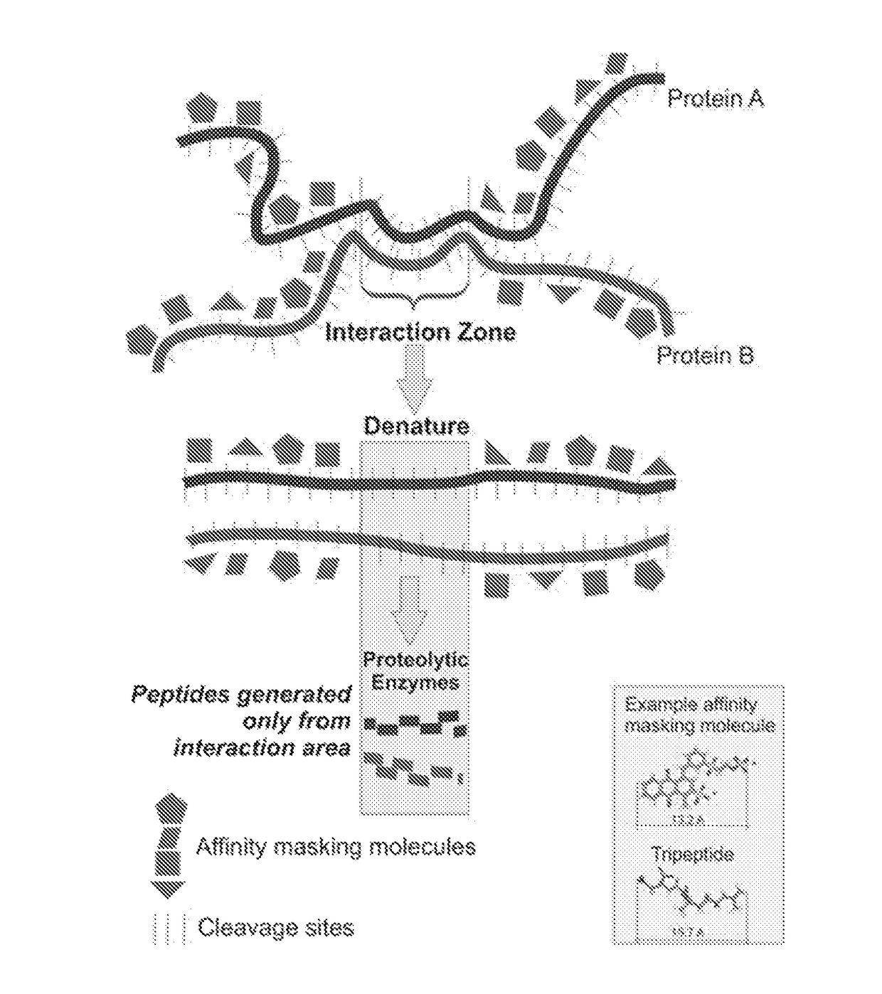
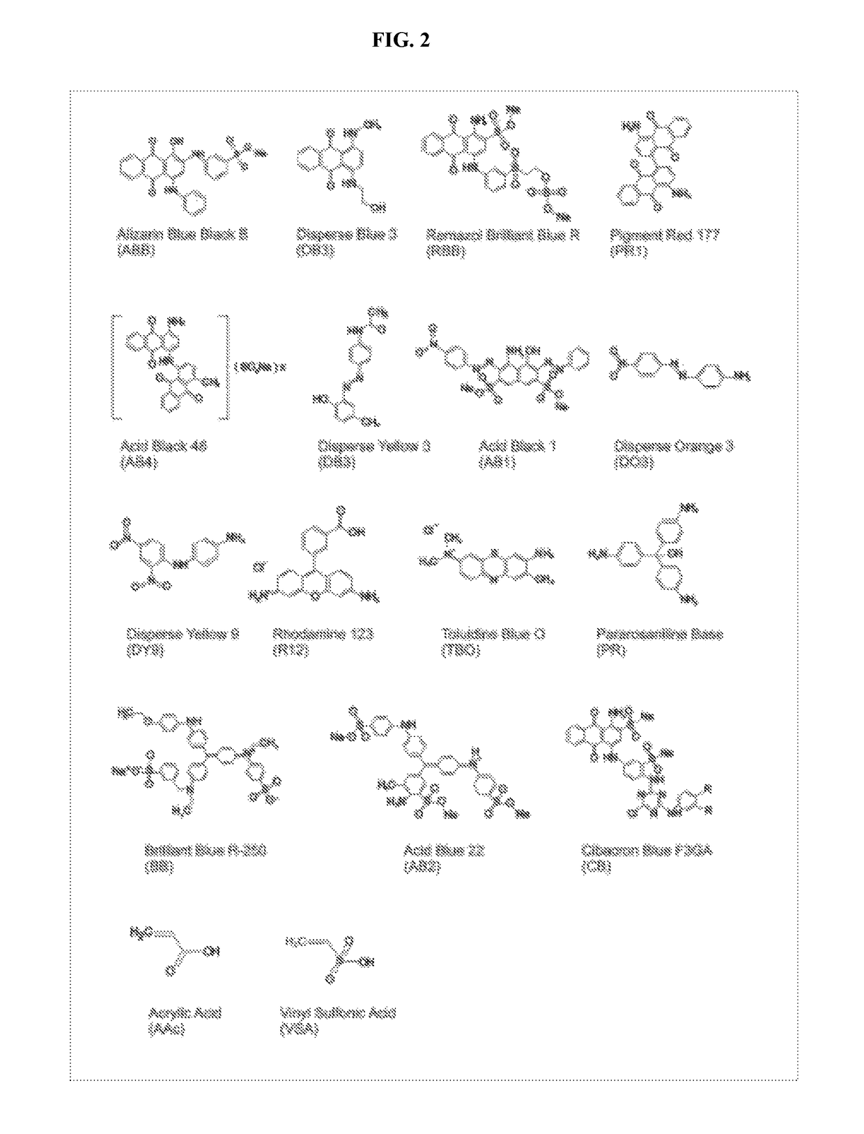
[0167]Masking paint molecules must be small enough in size so that the masking of the exposed surface of the individual protein molecule can be accomplished with adequate high resolution. After screening a numbers of molecules derived from dye chemistry, paints shown in Table 2 are an example of the types of molecules that can bind (with very high affinity) and mask protein domains with a resolution of approximately 3 amino acids. This provides sufficient resolution to effectively mask surface domains even over complex 3-D shape curvatures and can potentially achieve a masking density to cover all (trypsin or other protease) cleavage sites in any given protein. The instant paint molecules are ten to one-hundred times smaller than affinity peptides described in the literature. Protein peptides, that bind larger proteins, can not be used for the present technology because of their large size, much lower resolution of coating, susceptibility to proteinases, and lowe...
example 2
f Masking Affinity Molecules to the Surface of Proteins Blocks Trypsin Recognition Sites and Masks Trypsin Generation of Cleavage Fragments
[0177]Methods: Bovine serum albumin (BSA), aprotinin, and carbonic anhydrase II (1 mg / mL in PBS) were mixed with 10 molar excess disodium 1-amino-9,10-dioxo-4-[3-(2-sulfonatooxyethylsulfonyl) anilino] anthracene-2-sulfonate (RBB FIG. 4). Protein solutions were prepared in parallel with the same concentration without adding RBB, as controls. The solutions were immediately passed through a Sephadex column (PD MiniTrap G 25, GE Healthcare) and denatured with 8 M urea, reduced with 1 M dithiothreitol, alkylated with 0.5 M iodoacetamide, digested with trypsin, and subjected to mass spectrometry analysis (LTQ Orbitrap, Thermo Scientific).
[0178]Results: Comparison of the MS derived amino acid sequences indicated that the masking molecules effectively blocked trypsin cleavage and over multiple regions of the proteins as shown below the top sequence (1M) ...
example 3
on of the Masking Molecule(s) to Directly Sequence Interface Contact Areas Between Interacting Proteins
[0180]The IL1beta receptor and its protein ligand were chosen to demonstrate a utility of the present technology. Three dimensional structure of this complex is known by X ray crystallography (Nature, 386, 190-194, 1997).
[0181]Methods: The receptor protein and ligand protein were purchased from Adipogen and Biolegend, respectively. A solution containing the receptor protein and ligand was prepared (0.05 mg / mL in PBS) and placed at 37 C for 1 hour to allow binding between the two molecules to take place. The affinity masking molecule disodium 1-amino-9,10-dioxo-4-[3-(2-sulfonatooxyethylsulfonyl) anilino] anthracene-2-sulfonate (RBB) was added at a 10:1 molar excess. In order to separate the excess unbound masking molecule, the solution was immediately passed through a Sephadex column (PD MiniTrap G 25, GE Healthcare). The complexed proteins coated with the masking molecules were den...
PUM
| Property | Measurement | Unit |
|---|---|---|
| total length | aaaaa | aaaaa |
| concentrations | aaaaa | aaaaa |
| concentrations | aaaaa | aaaaa |
Abstract
Description
Claims
Application Information