

Ultra-sensitive multi-target lateral flow molecular assay with field-induced precipitation
a lateral flow molecular assay and ultra-sensitive technology, applied in the field of concentrating, separating, and quantifying biomolecules, can solve the problems of poor selectivity, false positive signals, and significant slowdown of throughput in many current assays, and achieve the effects of lowering g, lowering energy barrier, and raising initial energy
- Summary
- Abstract
- Description
- Claims
- Application Information
AI Technical Summary
Benefits of technology
Problems solved by technology
Method used
Image

Examples
example 1
[0099]Materials:
[0100]Fluorescein sodium salt was obtained from Fisher Scientific. Sodium chloride, sodium citrate and chloroauric acid trihydrate were obtained from Sigma-Aldrich. SYBR green I was received at 10,000× concentration from Invitrogen. Buffers were prepared by dilution from 10×PBS (pH 7.4) and 50×TAE (pH 8.4) obtained from Boston Bioproducts and 150 mM sodium phosphate buffer (pH 7.2) obtained from Teknova. Agarose gels were prepared at 1 wt % in 1×TAE using agarose powder from Ominpur and stored as liquids inside an oven maintained at 65° C. All agarose gels containing fluorescein were prepared in a like manner with the fluorescein concentration equal to 10 mM. QuikCast polyurethane casting resins (side A and side B) were obtained from TAP Plastics Inc. Acrifix 1R 0192 UV reactive cement was obtained from Evonik Industries while Loctite 3492 light cure adhesive was obtained from Loctite Corporation. RALEX cation-exchange membranes whose fixed negative charge is supplie...
example 2
[0107]Separation and Detection Protocol:
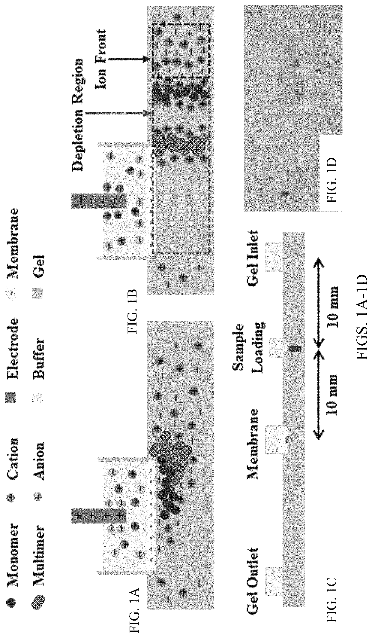
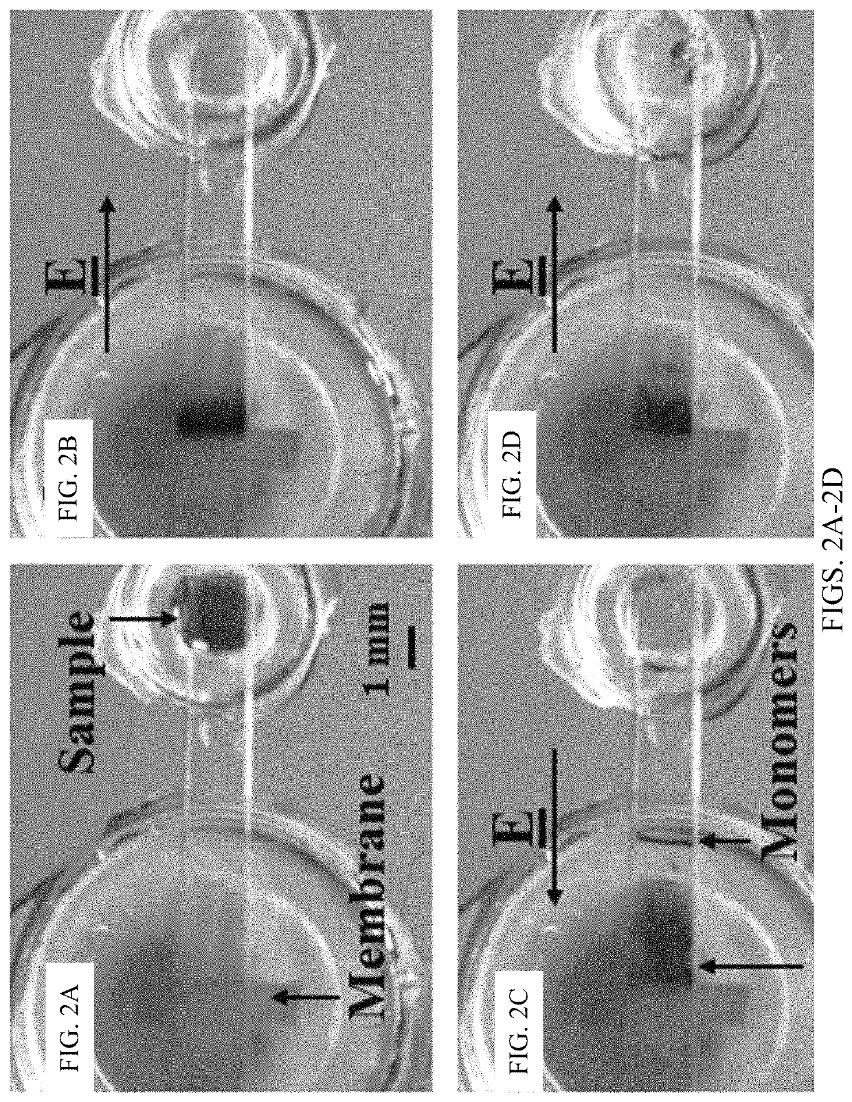
[0108]Chips, as shown in FIG. 1C, were filled with agarose gel and used after the gel solidified. The gel occupying the sample reservoir was removed and filled with 2 mL nanoparticle / DNA sample. The fluid reservoirs were all filled with 1×TAE buffer. Gel electrophoresis was conducted using a Keithley 2400A Sourcemeter with platinum electrodes as the voltage source. This example protocol consisted of four steps: enrichment, depletion, expulsion, and repacking. First, during enrichment, shown in FIG. 1A, the positive electrode was placed inside the membrane reservoir while the ground electrode was placed inside the inlet reservoir, and the sample was electrophoretically driven towards the membrane for five minutes by a 150V potential. The particles required approximately one minute to reach the membrane at which point the sample reservoir was refilled with agarose gel. Second, during depletion, typically eight to nine minutes, the field was reve...
example 3
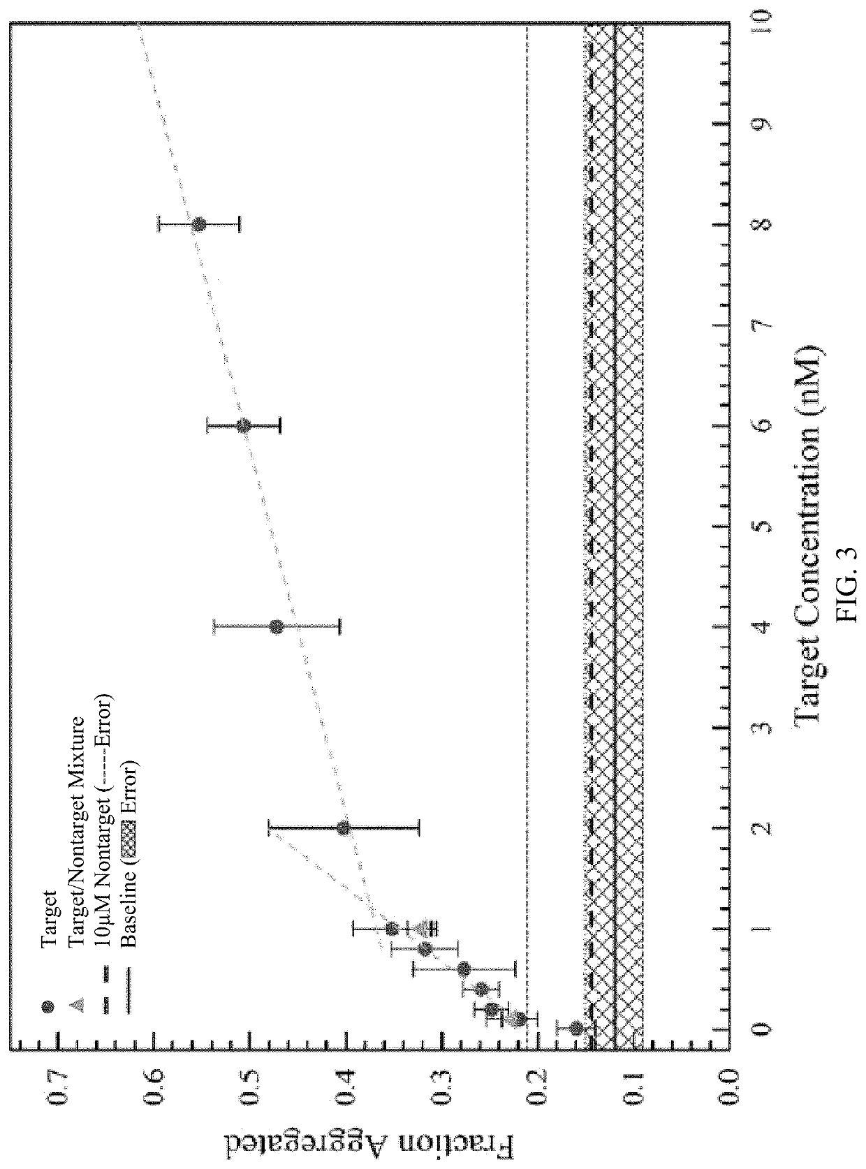
[0115]The selectivity of the sensor using targets with two base mismatches on each probe was examined (FIG. 4). The target sequence became 5′ / CTACCGTAGGACACCTGCCTCTTCCTCTTCCCTTCAAAAATAGCCCTAAAGCT ATTTCGG GCAACCA / 3′ (SEQ ID NO: 6). The experiments were carried out using slightly altered conditions where the distance between the channel inlet and membrane reservoirs was reduced to 15 mm, the enrichment time was increased to six minutes, and the applied potential during depletion underwent a step change. The maximum output of the sourcemeter, −200 V, was applied for one minute, and then the potential was lowered for the remainder of the depletion step (i.e. until the monomer reached the sample reservoir). The greatest selectivity occurred when the potential was lowered to 100 V and the target to mismatch signal ratio was greater than 3 (FIG. 4). These results were comparable to other assays such as the NanoBioArray chip based on hybridization of target-conjugated nanoparticles to surfa...
PUM
| Property | Measurement | Unit |
|---|---|---|
| diameter | aaaaa | aaaaa |
| diameter | aaaaa | aaaaa |
| voltages | aaaaa | aaaaa |
Abstract
Description
Claims
Application Information