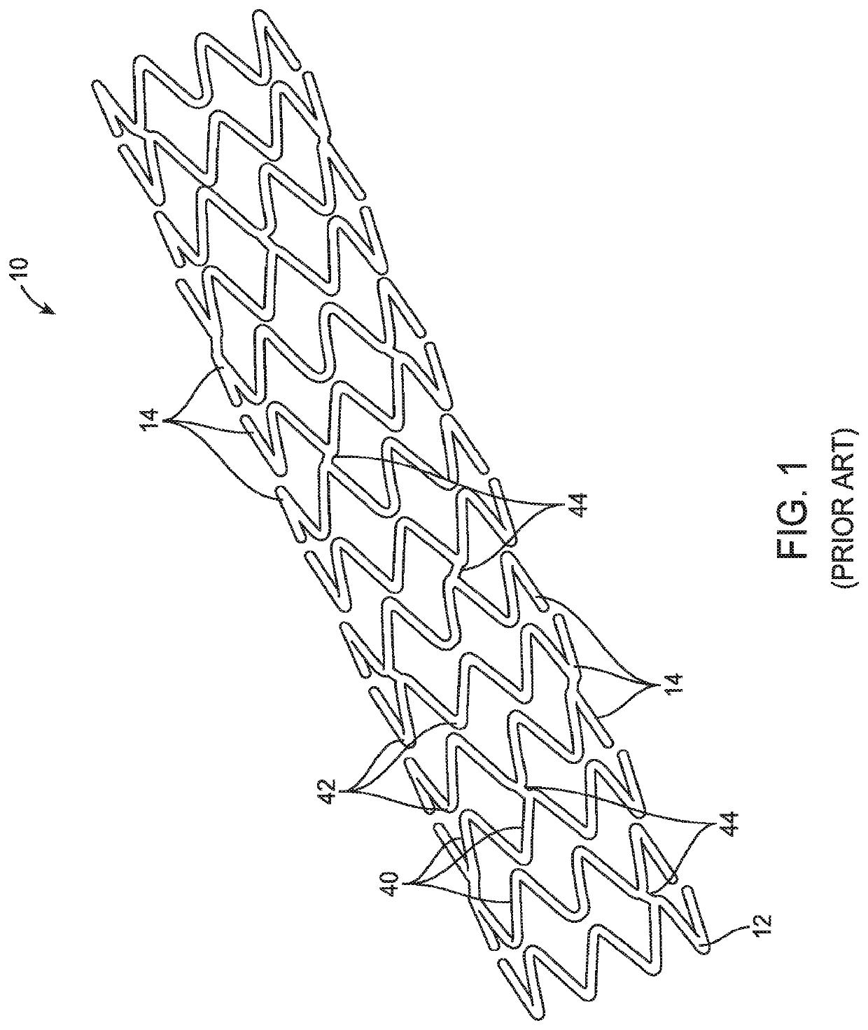
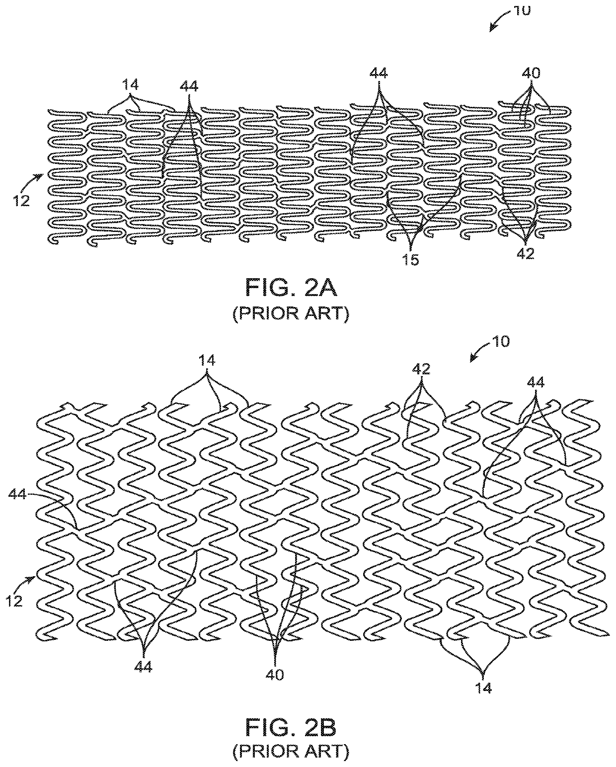
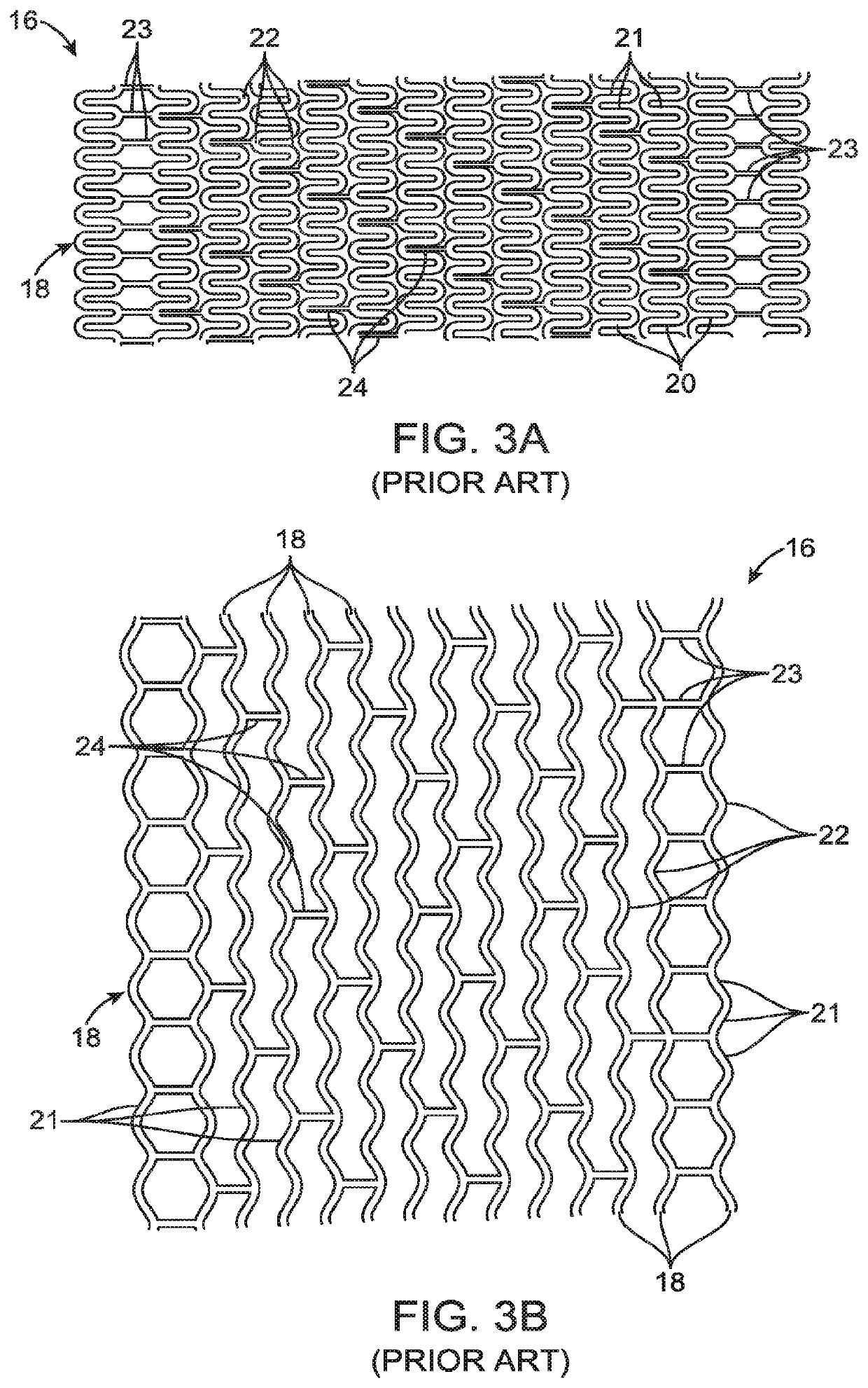
Uncaging stent

a stent and stent technology, applied in the field of uncaging stents, to achieve the effect of enhancing the structural integrity of the scaffold, less stress, and less stress

Active Publication Date: 2021-02-16
ELIXIR MEDICAL CORP
View PDF445 Cites 2 Cited by
  • Summary
  • Abstract
  • Description
  • Claims
  • Application Information

AI Technical Summary

Benefits of technology

The patent describes a stent that has rings without terminal ends or with very few terminal ends. This design has several benefits for the patient, including better vessel healing, reduction of irritation, and improved manufacturing. Additionally, the stent can have one or more separation regions that expand it to accommodate changes in tissue geometric configuration, which helps prevent blood leaks between the stent and adjacent tissue.

Problems solved by technology

Alternatively, as the scaffold is expanded, the axial links may undergo a deformation which is not sufficient to “unlock” their locked configuration, but after expansion in a physiologic environment, the axial links separate.

Method used


Image

  • Uncaging stent
  • Uncaging stent
  • Uncaging stent

Examples


example 1

[0885]Vasomotion evaluation by IVUS: A porcine animal having a control non-degradable plastically deformable stent (DESyne, Elixir Medical Inc. n=2) and a test stent of (PR153RG, n=4) having a 6-crown 3-link non-degradable plastically deformable stent patterned to have three evenly spaced separation regions per ring, with the S shaped axial links connecting adjacent rings was tested and followed up at the 3 month time point. The test devices were coated with a fast degrading (from 1-3 months) lactide copolymer covering the separation regions (including the inner, sides, and outer surfaces of the separation regions and the space within the separation regions) and covering the stent surfaces (luminal, abluminal and two side surface). The stents were further coated with a top coat of Novolimus and a lactide copolymer drug matrix. The abluminal and luminal coating thickness was about 10 microns. The test stents and the control stents (DESyne, Elixir Medical Inc.) were implanted in the c...

example 2

[0890]Finite Element Analysis Model testing for the different Tests listed below were conducted on the stent of the present invention. The mock artery had a wall thickness of 0.010″, regardless of diameter, and an inner diameter match the outer diameter of the stent. The artery wall material was modeled as a linear elastic material with a Poisson's ratio of 0.45, and exhibits a 5% diametric compliance when pressurized with 100 mmHg on the inner surface of the artery. The Young's modulus varied between 300 and 700 PSI, depending on the arterial diameter. The Stent material was a linear elastic material having a Young's modulus of between 36,000,000 and 39,000,000 PSI (36e6 to 39e6 PSI) and a Poisson's ratio of 0.3. FEA model used FEMAP and Nastran software and consisted of arrangements of 10 node modified Tet elements at a density shown to give results accurate to less than + / −5%. The minimum mesh density used in this analysis had 1 layer of elements through the thickness of the arte...

example 3

[0891]Three point Bend Test FIG. 15: A stent was patterned from Stent Material having a thickness of 75 microns, having a serpentine ring with each ring having 3 separation regions. The stent was in an expanded configuration of diameter 3.5 mm and is connected to a mock artery. Fixtures were connected at each end and in the middle. The minimum separation between the end connectors was 11 mm. The fixture at one end of the model was allowed to pivot along a line as shown below, but was otherwise fixed against movement. The fixture at the opposite end could pivot and move relative to the first end in the direction of the axis of the stent. The fixture on top in the middle was then displaced by 1 mm toward the stent, bending the stent and mock artery. Linear elastic analysis was used, resulting in an estimated Force of 1.4N prior to the formation of discontinuities, which dropped to 0.4N after formation of discontinuities. The stent of the current invention with discontinuities in the s...


PUM


Abstract

A stent (scaffold) or other luminal prosthesis comprising circumferential structural elements which provide high strength after deployment and allows for scaffold to uncage, and / or allow for scaffold or luminal expansion thereafter. The circumferential scaffold is typically formed from non-degradable material and will be modified to expand and / or uncage after deployment.

Description

CROSS REFERENCE TO RELATED APPLICATIONS[0001]This application is a continuation of U.S. patent application Ser. No. 16 / 786,736, filed Feb. 10, 2020, which is a continuation of PCT Application PCT / US18 / 46561, filed Aug. 13, 2018, which claimed priority from provisional patent application Nos. 62 / 622,741, filed Jan. 26, 2018; 62 / 577,624, filed Oct. 26, 2017; 62 / 558,273, filed Sep. 13, 2017; and 62 / 544,682, filed Aug. 11, 2017, the full disclosures of which are incorporated herein by reference.[0002]This application is a continuation-in-part of U.S. patent application Ser. No. 17 / 005,946, filed on Aug. 28, 2020, which is a continuation of U.S. patent application Ser. No. 16 / 518,657, filed Jul. 22, 2019, the full disclosures of which are incorporated herein by reference.BACKGROUND OF THE INVENTION1. Field of the Invention[0003]Balloon angioplasty was introduced to open vessels, particularly blood vessels which have narrowed as a result of plaque progression or a heart attack. In success...

Claims


Application Information

Patent Timeline
Patent Type & Authority Patents(United States)
IPC IPC(8): A61F2/915A61F2/89A61F2/90A61F2/07A61F2/82A61F2/24
CPCA61F2/915A61F2/89A61F2/90A61F2/2442A61F2002/075A61F2002/825A61F2002/91558A61F2002/91566A61F2002/91575A61F2002/91591A61F2210/0004A61F2210/0014A61F2210/0076A61F2220/0033A61F2220/0091A61F2230/001A61F2230/0006A61F2230/0008A61F2230/0069A61F2250/0031A61F2250/0036A61F2250/0048A61F2250/0067A61F2250/0071A61F2/07A61F2002/067A61F2002/91583A61F2250/0039
Inventor SIRHAN, MOTASIMYAN, JOHNBHAT, VINAYAKPARASCHAC, JOSEPHCRYER, BRETTSERNA, BENJAMYN
Owner ELIXIR MEDICAL CORP
Why Eureka
Try Eureka