Method and system for calculating SUV normalization coefficient in a SPECT quantitative tomographic image

a tomographic image and spectral technology, applied in tomography, image enhancement, instruments, etc., can solve the problems of not accurately recording the dose of medicamentous injections of most spectral tumor imaging, bringing certain errors to the evaluation, and weighing in clinical practice, so as to avoid operating errors, improve the accuracy of suv normalization coefficient calculation, and enhance the effect of suv value calculation

Active Publication Date: 2021-11-30
ATOMICAL MEDICAL EQUIP FO SHAN LTD
View PDF8 Cites 0 Cited by
  • Summary
  • Abstract
  • Description
  • Claims
  • Application Information

AI Technical Summary

Benefits of technology

[0052]In terms of accuracy, the present invention avoids errors in operating and recording the drug injection dose from affecting the accuracy of the SUV value calculation, and at the same time, it can correct and compensate for some special factors such as bladder uptake, radioactive contamination of body surface, at the level of plain film images according to clinical use habits and the actual situation of patients. For example, a predefined area of invalid radioactive uptake is segmented to improve the accuracy of the SUV normalization coefficient calculation, further enhance the accuracy of the SUV value calculation.
[0053]In terms of reliability, since all the input data of the algorithm of the present invention are derived from the Dicom image data and file header information output by the SPETOMOGRAPHIC device, it can be read and operated fully automatically, without the need for manual operation and keeping separate records, so it has higher reliability and better traceability.

Problems solved by technology

At present, the main problems that still exist at the level of image quantification methods rest with the consistency of equipment from different manufacturers and the quality control (periodical calibration) during the use of equipment.
If this part of the dose cannot be accurately corrected in the SUV calculation, it will cause quantitative standards between different patients to have differentiation, and brings certain errors to the evaluation of multiple imaging results of the same patient.
However, due to the limitation of actual needs and working conditions, most of the SPECT tumor imaging does not accurately record the subject's medicamentous injection dose and weigh in clinical practice.
Therefore, in terms of feasibility, it is limited to some extent that the method similar to the PET imaging is used to measure the average activity concentration of the medicant injected into the subject's body and further calculate the SUV value.

Method used


Image

  • Method and system for calculating SUV normalization coefficient in a SPECT quantitative tomographic image
  • Method and system for calculating SUV normalization coefficient in a SPECT quantitative tomographic image
  • Method and system for calculating SUV normalization coefficient in a SPECT quantitative tomographic image

Examples


embodiment 1

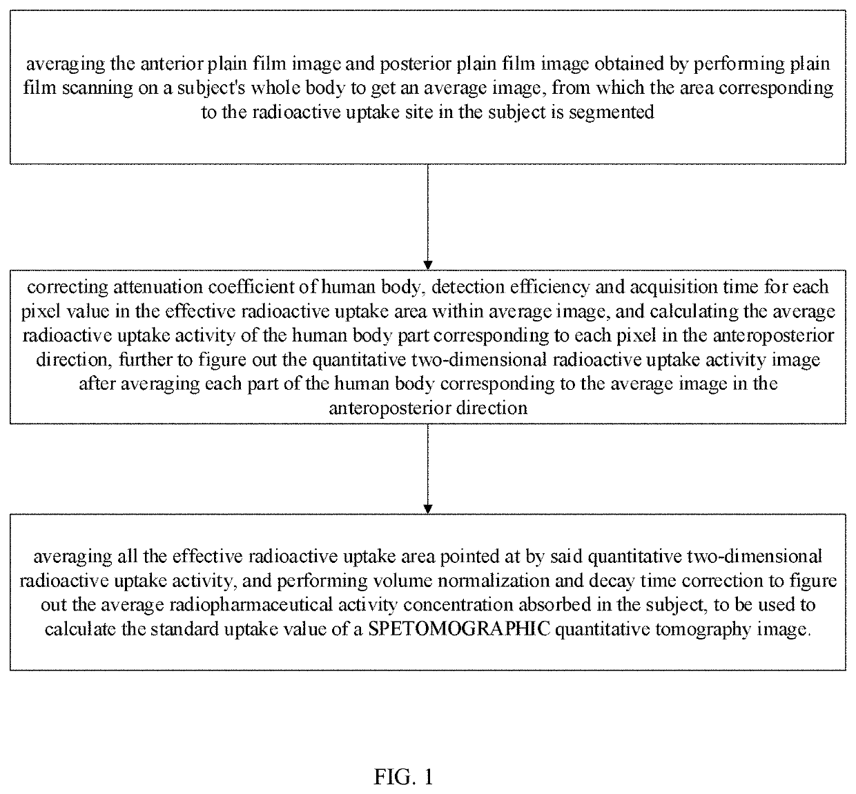
[0063]As shown in FIG. 1, the method for calculating SUV normalization coefficient in a SPETOMOGRAPHIC quantitative tomographic image in this embodiment includes the following steps:

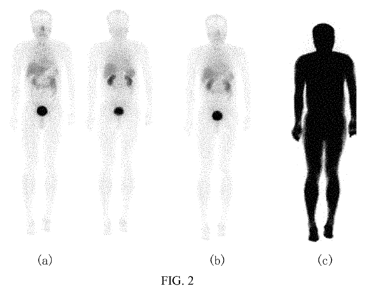
[0064]a) as shown in FIG. 2(a) and FIG. 2(b), averaging the anterior plain film image and posterior plain film image obtained by performing plain film scanning on a subject's whole body to get an average image, from which the area corresponding to the radioactive uptake site in the subject is segmented, and defined as an effective radioactive uptake area,

[0065]b) correcting attenuation coefficient of human body, detection efficiency and acquisition time for each pixel value in the effective radioactive uptake area within said average image, and calculating the average radioactive uptake activity of the human body part corresponding to each pixel in the anteroposterior direction, further to figure out the quantitative two-dimensional radioactive uptake activity image after averaging each part of the human...

embodiment 2

[0109]The system for calculating SUV normalization coefficient in a SPETOMOGRAPHIC quantitative tomographic image in this embodiment includes:

[0110]a module for calculating an effective radioactive uptake area, as shown in FIG. 2(a) and FIG. 2(b), which is used to average the anterior plain film image and posterior plain film image obtained by performing plain film scanning on a subject's whole body to get an average image, from which the area corresponding to the radioactive uptake site in the subject is segmented, and defined as an effective radioactive uptake area,

[0111]a module for calculating a quantitative two-dimensional radioactive uptake activity image, which is used to correct attenuation coefficient of human body, detection efficiency and acquisition time for each pixel value in the effective radioactive uptake area within said average image, and calculate the average radioactive uptake activity of the human body part corresponding to each pixel in the anteroposterior dir...

embodiment 3

[0145]This example applies the method described in Example 1 to two groups of clinical trial subjects where 99mTc-3PRGD2 is used for SPETOMOGRAPHIC imaging. The two groups of subjects come from different medical centers, the clinical center1 and the clinical center2, of which the subjects from the clinical center1 are the group of the patients who are clinically diagnosed with breast cancer, and the subjects from the clinical center2 are the group of the healthy volunteers. The SPETOMOGRAPHIC systemic plain film and the local SPETOMOGRAPHIC tomographic imaging of at least one site are performed on both groups of subjects, and the injection dose, injection time and subject weight are recorded before imaging. The correlation analysis is performed on the average radiopharmaceutical activity concentration absorbed in the subject which is figured out by using the method described in Embodiment 1, and the average concentration which is figured out by using the method that the injection do...


PUM


Abstract

The present invention discloses a method and system for calculating SUV normalization coefficient in a SPECT quantitative tomographic image, comprises the following steps: a) averaging the anterior plain film image and posterior plain film image obtained by performing plain film scanning on a subject's whole body to get an average image, from which the area corresponding to the radioactive uptake site in the subject is segmented, b) correcting attenuation coefficient of human body, detection efficiency and acquisition time for each pixel value in the effective radioactive uptake area within said average image, c) averaging all the effective radioactive uptake area pointed at by said quantitative two-dimensional radioactive uptake activity image obtained in step b, and performing volume normalization and decay time correction to figure out the average radiopharmaceutical activity concentration absorbed in the subject.

Description

CROSS-REFERENCE TO RELATED APPLICATIONS[0001]This application claims the benefit of priority from Chinese Patent Application No. 201910827035.5, filed on Sep. 3, 2019. The content of the aforementioned application, including any intervening amendments thereto, is incorporated herein by reference in its entirety.TECHNICAL FIELD[0002]The present invention relates to the technical field of nuclear medicine imaging, and more specifically to a method and system for calculating SUV normalization coefficient in a SPECT quantitative tomographic image.BACKGROUND ART[0003]The standardized uptake value (SUV) is a semi-quantitative indicator commonly used in the positron emission tomography (PET) in the tumor image analysis, and specifically refers to the ratio of the radioactivity concentration of the imaging agents absorbed by local tissues to the average radioactivity concentration injected systemically. The concentration ratio is shown in the following formula:[0004]SUV=r(a′⁢ / ⁢w)[0005]where...

Claims


Application Information

Patent Timeline
Patent Type & Authority Patents(United States)
IPC IPC(8): G06K9/00G16H30/20G06T7/11G06N3/02G06T7/00
CPCG16H30/20G06N3/02G06T7/0012G06T7/11G06T2207/10108G06T2207/20081G06T2207/20084A61B6/5211G06T2207/30004G06T2207/30196A61B6/037G16H30/40G16H50/20G06N20/00
Inventor CHEN, SI
Owner ATOMICAL MEDICAL EQUIP FO SHAN LTD
Why Eureka
Try Eureka