Interventional device recognition

a technology of interventional devices and ultrasound, applied in the field of ultrasound localization of interventional devices, can solve the problems of difficult visualization of interventional devices such as needles, catheters and surgical tools in ultrasound images, mechanical constraints of such instruments hamper the ability to position the ultrasound detector at will, and localization systems, etc., to achieve accurate determination and improve the determination of the position of the distal end

Active Publication Date: 2022-03-15
KONINKLJIJKE PHILIPS NV
View PDF10 Cites 0 Cited by
  • Summary
  • Abstract
  • Description
  • Claims
  • Application Information

AI Technical Summary

Benefits of technology

[0012]In so doing, an apparatus is provided which can be used to track the position of an interventional device within an ultrasound field. Depending on the particular type of interventional device that is being tracked, the geometry-providing unit selects the corresponding transducer-to-distal-end length, and indicates this in the reconstructed image in combination with the out-of-plane distance of the ultrasound transducer. Thus, the apparatus provides improved determination of the position of the distal end of the interventional device. Moreover, because both the transducer-to-distal-end length and the out-of-plane distance are indicated in the reconstructed image, the position of the distal end of the interventional device in relation to the image plane is more accurately determined.
[0015]In so doing the data stored in the data carrier of the interventional device, when received by the apparatus, brings about the benefit of improved determination of the position of the distal end of the interventional device respective the image plane of the beamforming ultrasound imaging system.

Problems solved by technology

Interventional devices such as needles, catheters and surgical tools are often difficult to visualize in an ultrasound image due to the specular nature of their reflectivity, particularly at unfavorable incidence angles.
A drawback of the above-mentioned systems arises from the fact that it is the position of the ultrasound detector that is determined and subsequently displayed in the ultrasound image.
However, the mechanical constraints of such instruments hampers the ability to position the ultrasound detector at-will, for example at the tip of a needle where it might interfere with insertion.
Another drawback of known localization systems occurs more specifically when they are used in conjunction with a planar ultrasound imaging system.
Owing to the separation between the ultrasound detector being tracked and the functional part on the instrument, the ultrasound detector may be out-of-plane when the functional part is in-plane, but poorly visible under ultrasound.

Method used


Image

  • Interventional device recognition
  • Interventional device recognition
  • Interventional device recognition

Examples


Embodiment Construction

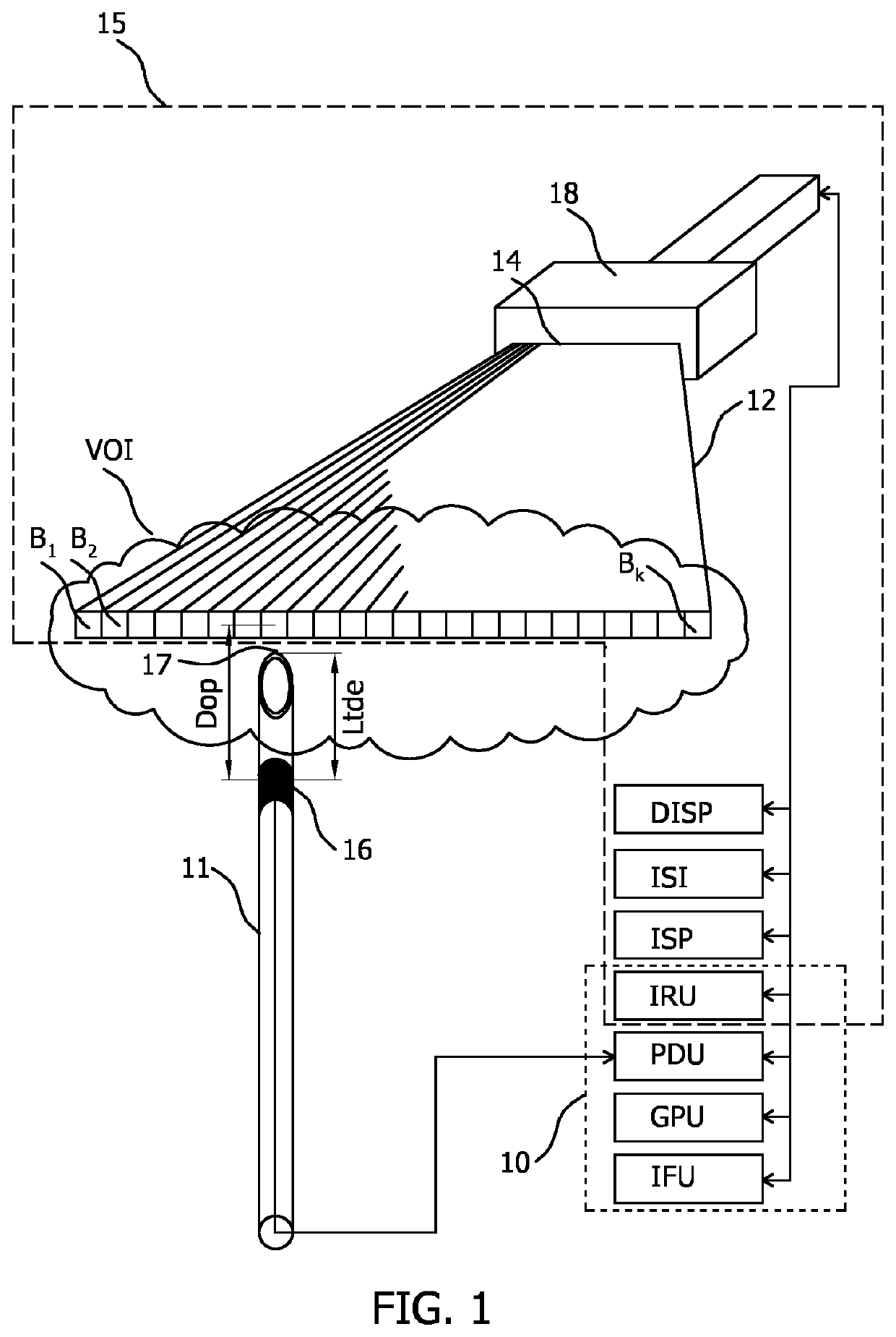
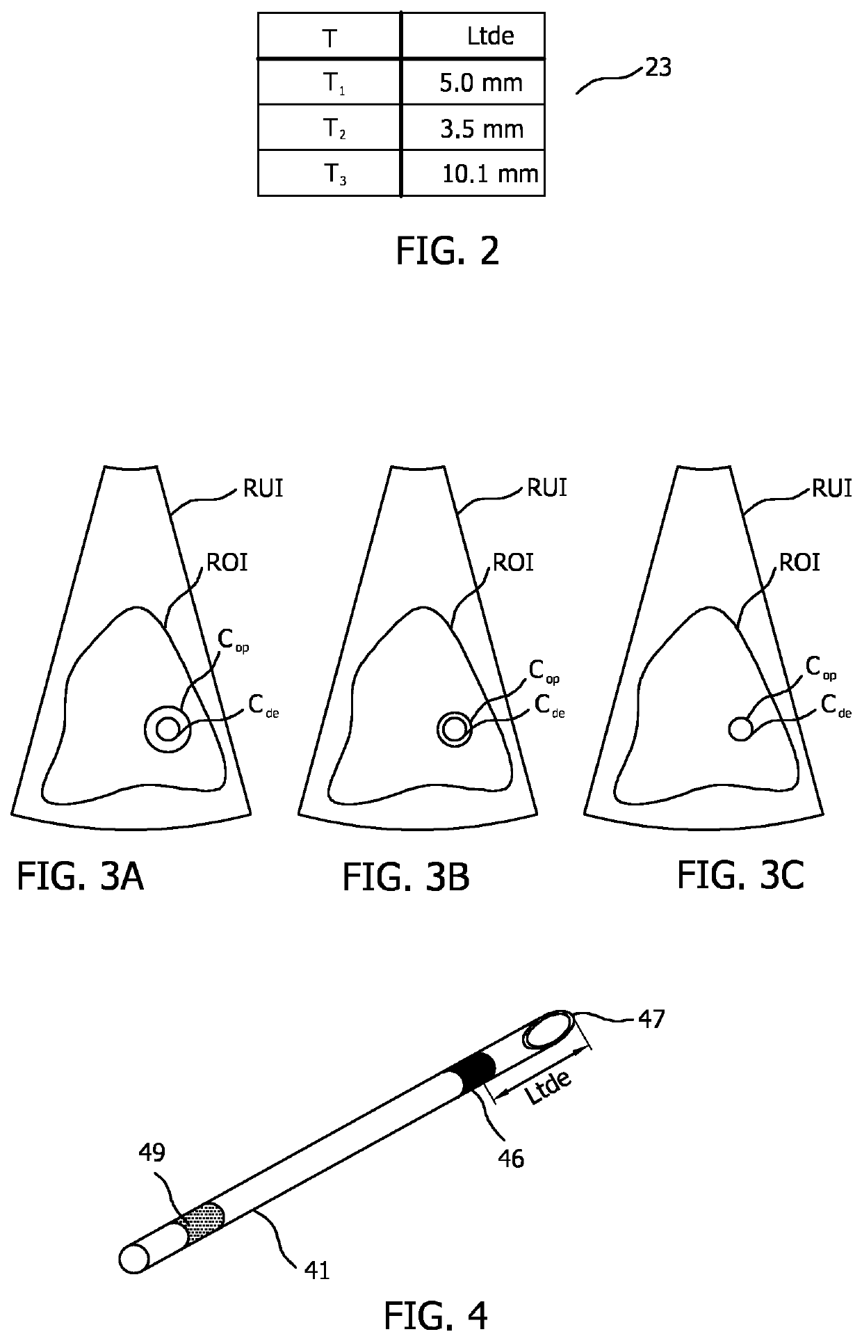
[0023]In order to illustrate the principles of the present invention, various systems are described in which the position of an interventional device, exemplified by a medical needle, is determined within the image plane of an ultrasound field defined by the beams emitted by the linear array of a 2D ultrasound imaging probe.

[0024]It is however to be appreciated that the invention also finds application in determining the positon of other interventional devices such as a catheter, a guidewire, a probe, an endoscope, an electrode, a robot, a filter device, a balloon device, a stent, a mitral clip, a left atrial appendage closure device, an aortic valve, a pacemaker, an intravenous line, a drainage line, a surgical tool such as a tissue sealing device or a tissue cutting device.

[0025]It is also to be appreciated that the invention finds application in beamforming ultrasound imaging systems having other types of imaging probes and other types of ultrasound arrays which are arranged to p...


PUM


Abstract

The present invention relates to an apparatus (10) for tracking a position of an interventional device (11) respective an image plane (12) of an ultrasound field. The position includes an out-of-plane distance (Dop). A geometry-providing unit (GPU) includes a plurality of transducer-to-distal-end lengths (Ltde1 . . . n), each length corresponding to a predetermined distance (Ltde) between a distal end (17, 47) of an interventional device (11, 41) and an ultrasound detector (16, 46) attached to the interventional device, for each of a plurality of interventional device types (T1 . . . N). An image fusion unit (IFU) receives data indicative of the type (T) of the interventional device being tracked; and based on the type (T): selects from the geometry-providing unit (GPU), a corresponding transducer-to-distal-end length (Ltde); and indicates in a reconstructed ultrasound image (RUI) both the out-of-plane distance (Dop) and the transducer-to-distal-end length (Ltde) for the interventional device within the ultrasound field.

Description

CROSS-REFERENCE TO PRIOR APPLICATIONS[0001]This application is the U.S. National Phase application under 35 U.S.C. § 371 of International Application Serial No. PCT / EP2016 / 079522, filed on Dec. 2, 2016, which claims the benefit of U.S. Patent Application No. 62 / 267,948, filed on Dec. 16, 2015 and European Patent Application No. 16154967.0, filed on Feb. 10, 2016. This application is hereby incorporated by reference herein.FIELD OF THE INVENTION[0002]The present invention relates to the localization of an interventional device using ultrasound. More specifically it relates to the recognition of the type of interventional device that is attached to an ultrasound-based localization system.BACKGROUND OF THE INVENTION[0003]Interventional devices such as needles, catheters and surgical tools are often difficult to visualize in an ultrasound image due to the specular nature of their reflectivity, particularly at unfavorable incidence angles.[0004]In one solution to this problem, U.S. Pat. ...

Claims


Application Information

Patent Timeline
Patent Type & Authority Patents(United States)
IPC IPC(8): G01S5/30A61B8/08G01S15/89A61B34/20A61B90/00A61B8/12G01S7/52A61B8/00A61B17/34A61B90/96A61B90/98
CPCG01S5/30A61B8/0841A61B8/12A61B8/4438A61B8/4488A61B17/3403A61B34/20A61B90/37G01S7/52026G01S7/52073G01S15/899G01S15/8915G01S15/8918G01S15/8977A61B8/463A61B90/96A61B90/98A61B2017/3413A61B2034/2063A61B2090/378A61B2090/3782A61B2090/3786
Inventor STAPERT, HENDRIK ROELOFSNIJDER, CARINAJAIN, AMEET KUMARDE WIJS, WILLEM-JAN AREND
Owner KONINKLJIJKE PHILIPS NV
Why Eureka
Try Eureka