Adenocarcinoma detection method

a technology of adenocarcinoma and detection method, which is applied in the direction of peptide sources, instruments, peptides, etc., can solve the problems of difficult early detection and difficult to diagnose lung adenocarcinoma in a fundamentally treatable stag

Active Publication Date: 2022-09-06
UNIVERSITY OF MIYAZAKI +1
View PDF12 Cites 0 Cited by
  • Summary
  • Abstract
  • Description
  • Claims
  • Application Information

AI Technical Summary

Benefits of technology

[0058]According to the present invention, adenocarcinoma can be detected by a minimally invasive method using body fluids such as urine and blood.

Problems solved by technology

Among them, the lung adenocarcinoma is one of adenocarcinomata of which initial symptom is unlikely to appear and of which early detection is difficult.
In the case of CEA, it is difficult to diagnose lung adenocarcinoma in a fundamentally treatable stage.
However, these markers are subjected to the conditions where blood is collected from patients, and thus are invasive.

Method used


Image

  • Adenocarcinoma detection method
  • Adenocarcinoma detection method
  • Adenocarcinoma detection method

Examples


example 1

[Example 1] Collection of Body Fluids

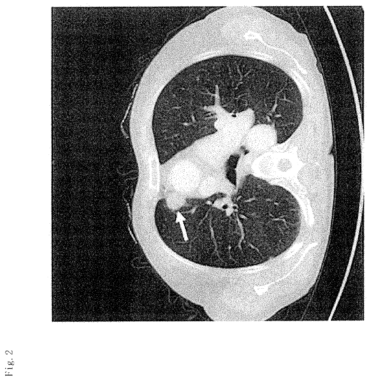
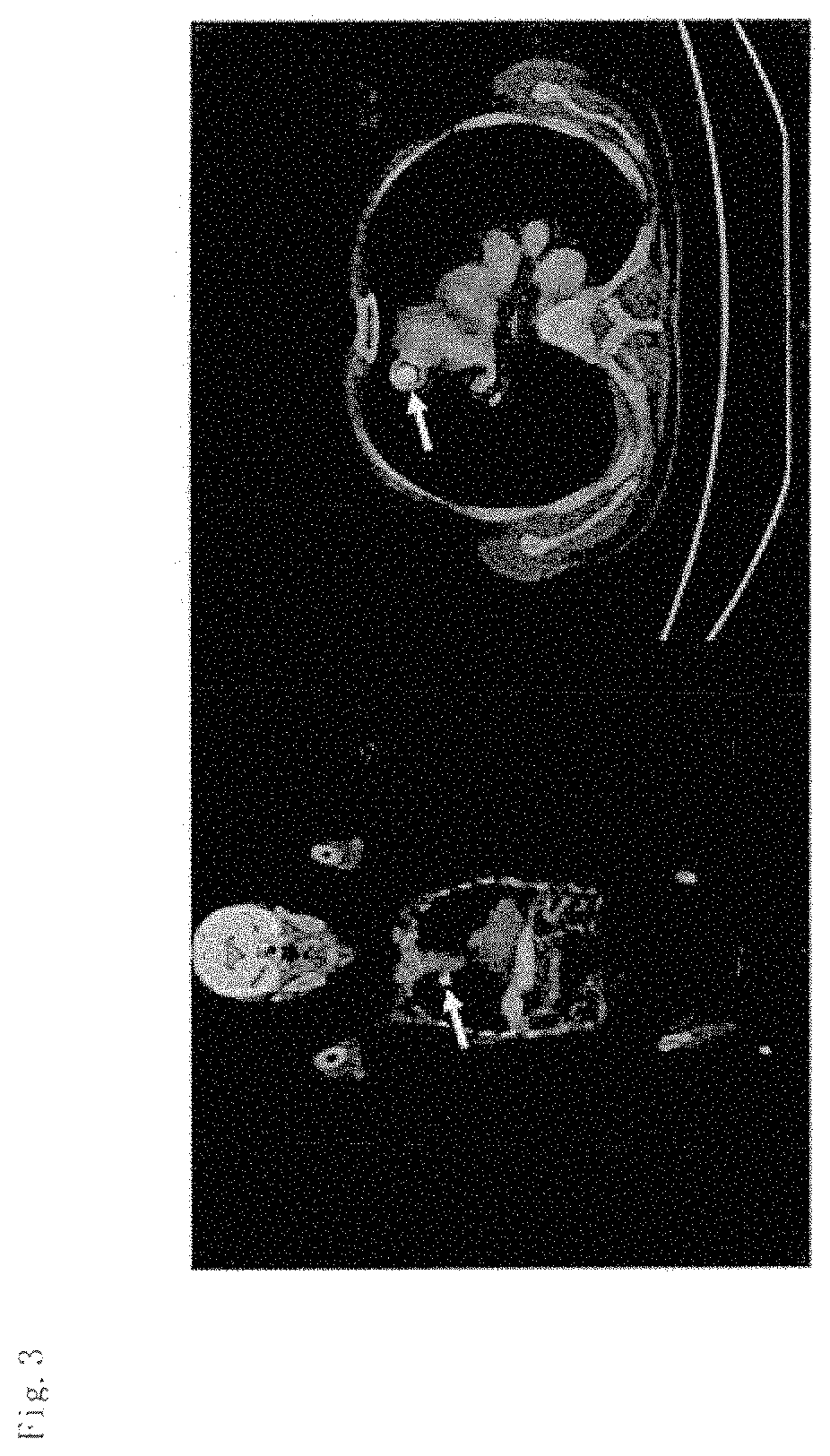
[0149]Among patients with pulmonary nodule shadows or tumor shadows, suspected of primary lung cancer by chest image examination, patients diagnosed with lung adenocarcinoma were selected by surgery, transbronchial biopsy, lymph node biopsy or cytology. Early morning midstream urine was collected from selected lung adenocarcinoma patients and healthy individuals using sterile cups. The collected urine samples were stored at −80° C. until analysis.

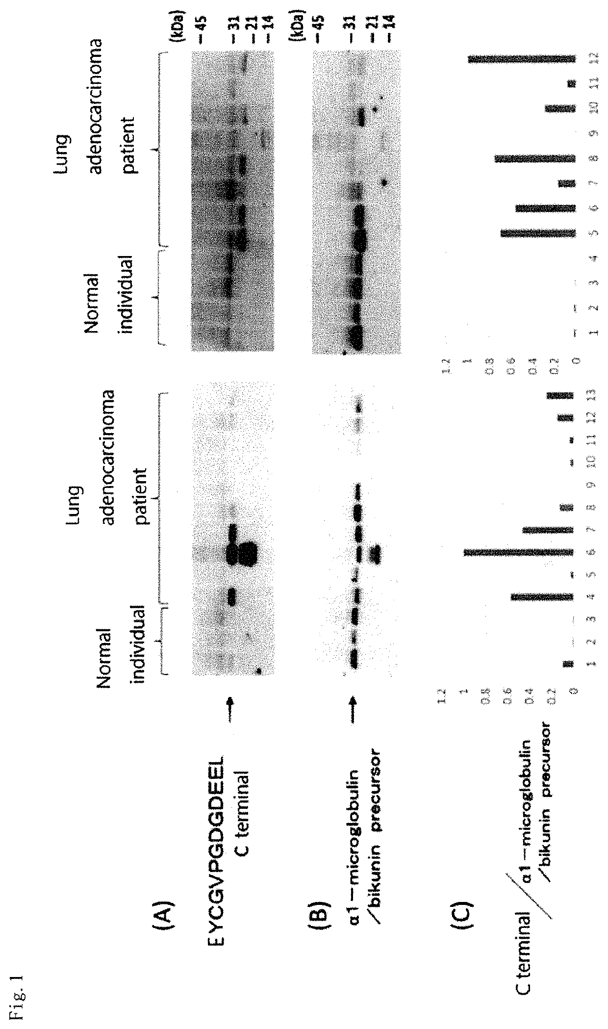
[0150]C-terminal protein fragments in urine from 85 cases with lung adenocarcinoma and 25 healthy individuals were comprehensively analyzed. A breakdown of clinical stages of the cases with lung adenocarcinoma showed that 18 cases were in clinical stage IA, 5 cases were in clinical stage IB, 1 case was in clinical stage IIA, 0 case was in clinical stage IIB, 6 cases were in clinical stage IIIA, 5 cases were in clinical stage IIIb, and 50 cases were in clinical stage IV. The clinical stage was judged acco...

example 2

[Example 2] Detection by Mass Spectrometry

[0151]Urine samples (˜50 mL) were collected and markers were searched by the procedures of pretreatment, acquisition of analytical data, and statistical analysis. In the pretreatment, urine samples were concentrated to 200 to 250-folds using Amicon Ultra-15 (10 kDa molecular weight cut-off) and Amicon Ultra-4 (10 kDa molecular weight cut-off) (Merck KGaA) and washed 3 times with 3 mL of triethylammonium hydrogencarbonate solution containing 100 mM NaCl, thereby removing low molecular weight molecules. Thereafter, concentrated specimens were obtained. The proteins in the concentrated specimens were quantified. The concentrations of all the specimens were adjusted to 10 mg / mL of total protein using a buffer solution and the resulting specimens were used in the subsequent analysis process. Subsequently, the samples were subjected to reductive alkylation and then digested with trypsin in a buffer prepared with a constant concentration of H218O (...

example 3

[Example 3] Selection of Markers

[0152]Regarding the peak intensity (Example 2) of each of the C-terminal trypsin-digested peptides of the MS / MS spectra obtained from urine samples from lung adenocarcinoma patients and healthy individuals based on the statistical analysis, the relative ratio of the control specimen to the peak intensity was calculated, and comparison and evaluation were made between lung adenocarcinoma patients and healthy individuals. Mann-Whitney's U test was used as the assay method. For statistical analysis, JMP12 (SAS Institute Inc, Cary, N.C.) was used. The peak intensities of protein fragments in urine were compared between 85 cases with lung adenocarcinoma or 24 cases with early-stage lung adenocarcinoma and healthy individuals. The protein fragments satisfying the criteria that the ROC-AUC value was 0.6 or more or the p value was less than 0.1 were used as marker candidates. A total of 18 types of protein fragments were identified as lung adenocarcinoma diag...


PUM

PropertyMeasurementUnit
concentrationaaaaaaaaaa
molecular weightaaaaaaaaaa

Abstract

There is provided a simple and minimally invasive adenocarcinoma detection method. The adenocarcinoma detection method of the present invention includes a step of detecting in vitro a presence or absence of an abnormal cleavage in a specific protein in a test subject-derived sample. The abnormal cleavage in the specific protein is, for example, a cleavage resulting in one or more breaks in a peptide bond in the specific protein and / or a cleavage resulting in a deletion of one or two more amino acid residues at one or more sites of the specific protein. The adenocarcinoma detection method of the present invention includes a step of detecting a presence or amount of a protein having the abnormal cleavage or a decrease in an amount of a normal protein.

Description

CROSS-REFERENCE TO RELATED APPLICATIONS[0001]This application is a national stage entry under 35 U.S.C. § 371 of International Application No. PCT / JP2017 / 005729, filed Feb. 16, 2017, which claims priority to Japanese Application No. 2016-030267, filed Feb. 19, 2016, the content of each of which is incorporated by reference in its entirety.SEQUENCE LISTING[0002]The instant application contains a Sequence Listing which has been submitted electronically in ASCII format and is hereby incorporated by reference in its entirety. Said ASCII copy, created on Jun. 8, 2020, is named 104788-0320_SL-2.txt and is 51,647 bytes in size.TECHNICAL FIELD[0003]The present invention relates to an adenocarcinoma detection method.BACKGROUND ART[0004]The adenocarcinoma is one of epithelial malignant tumors arising from cells in the secretory gland tissue. Lung adenocarcinoma, liver adenocarcinoma, pancreatic adenocarcinoma, lymphatic adenocarcinoma, uterine adenocarcinoma, seminal adenocarcinoma, gastric a...

Claims


Application Information

Patent Timeline
Patent Type & Authority Patents(United States)
IPC IPC(8): G01N33/574C07K14/705C07K14/78G01N33/68
CPCG01N33/57423C07K14/705C07K14/78G01N33/6803C07K16/18C07K16/28C07K14/7056C07K14/81C07K14/4726C07K14/4702G01N33/6818
Inventor NAKAZATO, MASAMITSUMATSUMOTO, NOBUHIROARIMURA, YASUJITSUBOUCHI, HIRONOBUTAKAO, TOSHIFUMIOKUMURA, NOBUAKIFUKUDA, MASAFUMI
Owner UNIVERSITY OF MIYAZAKI
Why Eureka
Try Eureka