

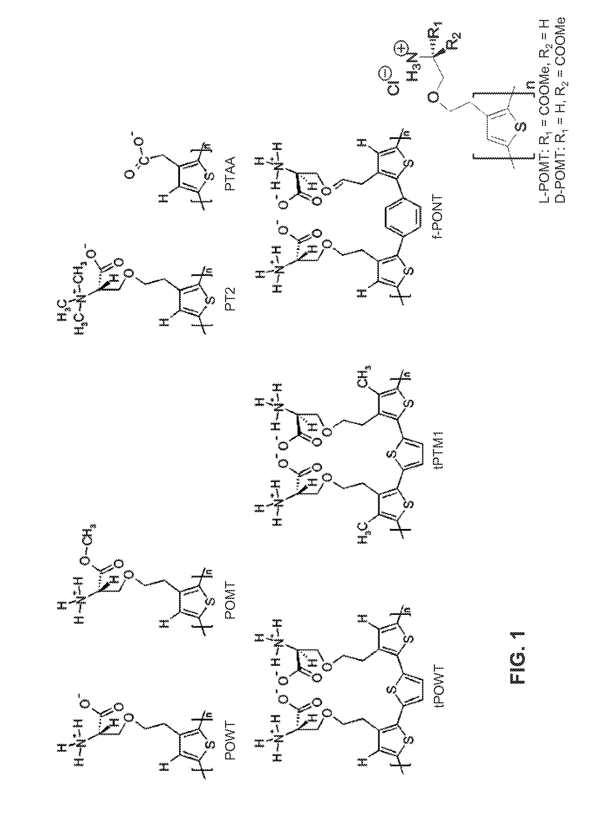
Binding of pathological forms of proteins using conjugated polyelectrolytes
a technology of conjugated polyelectrolytes and proteins, applied in the field of use, can solve the problems of inability to separate, use of antibodies as capture agents, harmful or even toxic, and inability to function, and achieve enhanced selectivity, enhanced affinity, and facilitate capture
- Summary
- Abstract
- Description
- Claims
- Application Information
AI Technical Summary
Benefits of technology
Problems solved by technology
Method used
Image

Examples
example 1
Capture and Detection of PrP-Amyloid in Solution Using Conjugated Polyelectrolytes
[0099]Surface detection by means of fluorescence microscopy PrP and PrP-amyloid sample, the latter being PrPSc- or PrPres-like, is contained in a test tube. PrP-amyloid was generated through dialysis of 1 mg / ml recHuPrP90-231 dissolved in 3 M guanidinium hydrochloride versus water. Another protocol to generate PrP-amyloid is by shaking in a test tube at a high salt concentration. Adding the conjugated polyelectrolyte PTAA (10 μg / ml) to this solution, containing PrP and PrP-amyloid, causes PTAA to capture, bind and stain as well, PrP-amyloid. The PTAA / PrP-amyloid formation can then be left to sediment to the bottom of the test tube, and can then easily be isolated from the sample and detected if desired. Other ways to collect PTAA / PrP-amyloid is, but not limited to, filtration, centrifugation, capture on a hydrophobic surface, capture on a surface with covalently attached PTAA, using functionalized part...
example 2
[0100]Filtration, Capture
[0101]In order to separate native and fibril insulin and hence purifying the fibril insulin, filtration of polymer-insulin samples were performed. Samples tested were native insulin and fibril insulin, with and without PTAA. Initial PTAA concentration were 0.5 mg / ml and insulin concentration 2 mg / ml, and samples contained 10 μl PTAA+25 μl insulin to 1 ml in phosphate buffer 20 mM pH 8. For all samples fluorescence measurements were performed both before and after filtration, and the filters were opened and visually inspected. Filters used were 0.22 μm Millex-gu, and 1 ml of every sample were filtered through the filters.
[0102]The fluorescence spectra of PTAA-native insulin (PTAA-ins) and PTAA-fibril insulin (PTAA-fib) are shown in FIG. 9 before and after filtration through the 0.22 μm filter. Before filtration the spectra look as expected. After filtration it is obvious that samples with PTAA and fibril insulin is retained in the filter (seen as a decrease i...
example 3
Capture and Detection of Insulin-Amyloid in Solution Using Conjugated Polyelectrolytes. Solution Detection by Means of Fluorescence Measurements
[0106]Samples with native insulin containing 0, 1, 5, 50 and 100% fibrils (2 mg / ml, 25 μl) are mixed with PTAA (0.05 mg / ml, 10 μl) and 965 μl 20 mM phosphate buffer pH 7.0 in a test tube. Fibril insulin was generated through incubation of native insulin at 65 degrees Celsius at pH 2 for 8 hours. The samples were centrifuged at 10000 rpm for 5 minutes, the supernatant was removed, new buffer was added, and the procedure was repeated once. The PTAA binds to the fibril insulin and sediments to the bottom of the test tube under the centrifugation. The samples were then measured using a fluorescence microplate reader. Reference samples of PTAA-native insulin and PTAA-fibril insulin that had not been centrifuged were also measured. The results are shown in FIG. 12.
[0107]The PTAA-fibril aggregates sediments to the bottom of the test tube and can ea...
PUM
| Property | Measurement | Unit |
|---|---|---|
| wavelengths | aaaaa | aaaaa |
| concentration | aaaaa | aaaaa |
| concentration | aaaaa | aaaaa |
Abstract
Description
Claims
Application Information