

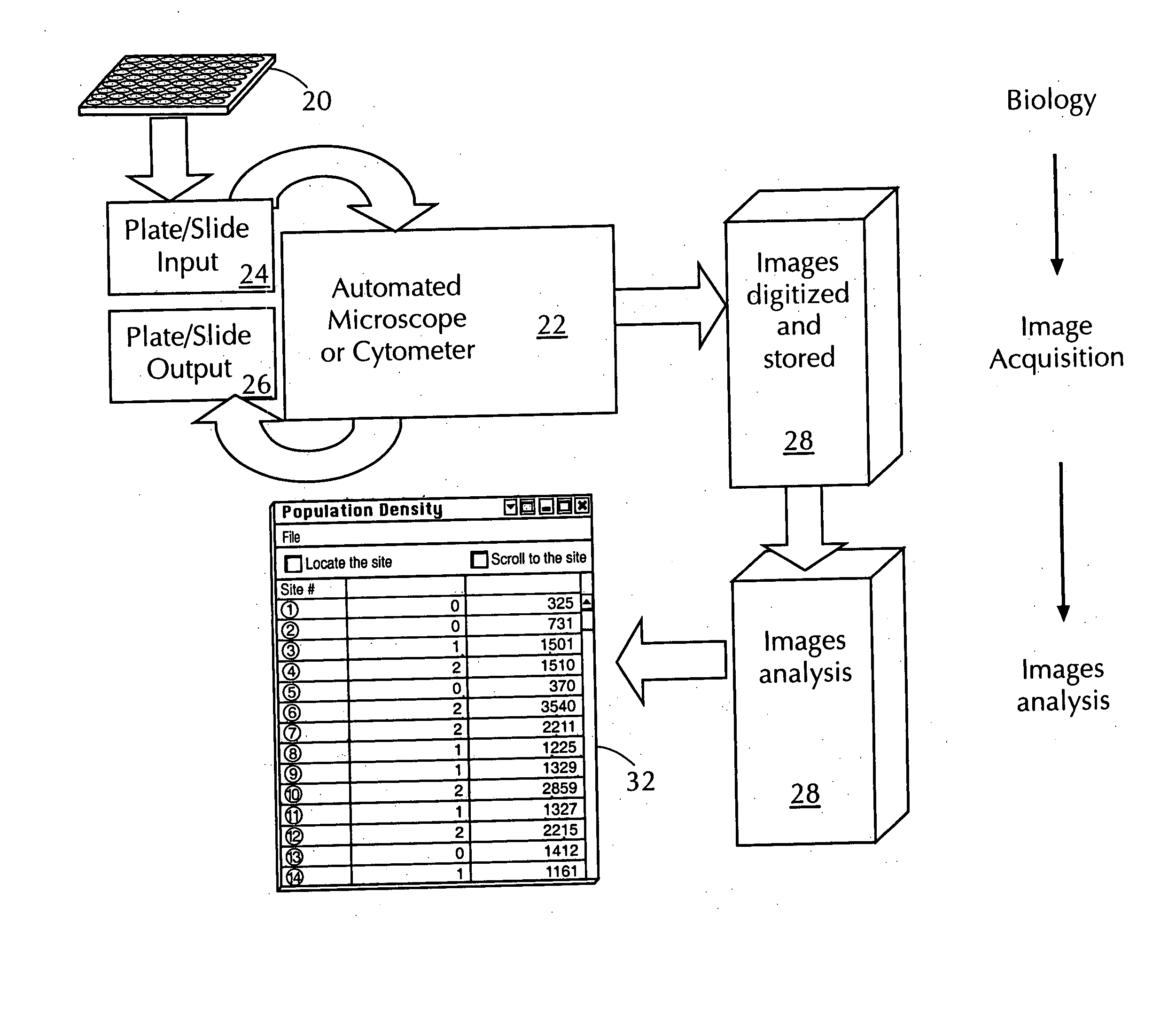
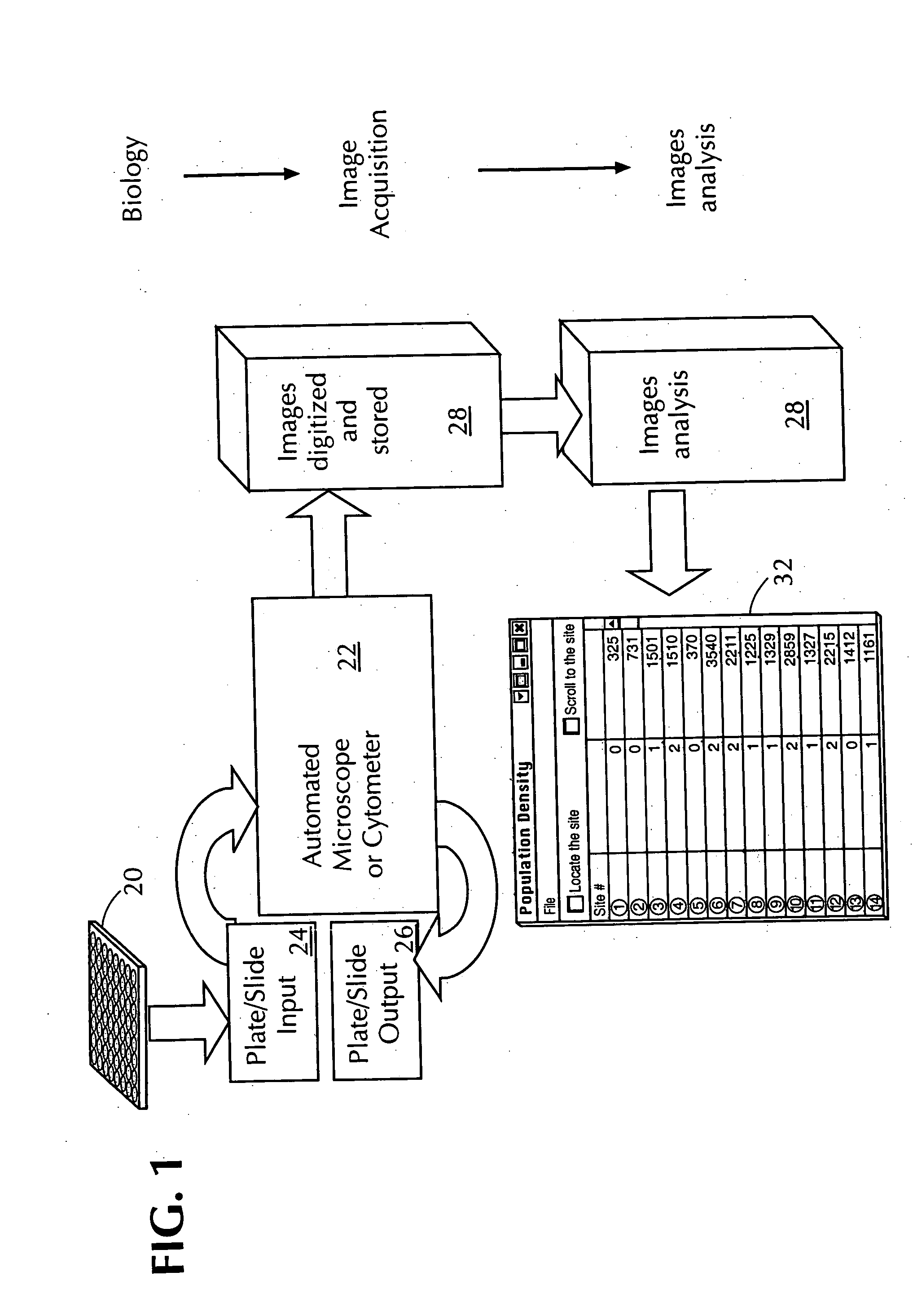
Automated in vitro cellular imaging assays for micronuclei and other target objects
- Summary
- Abstract
- Description
- Claims
- Application Information
AI Technical Summary
Benefits of technology
Problems solved by technology
Method used
Image

Examples
example 1
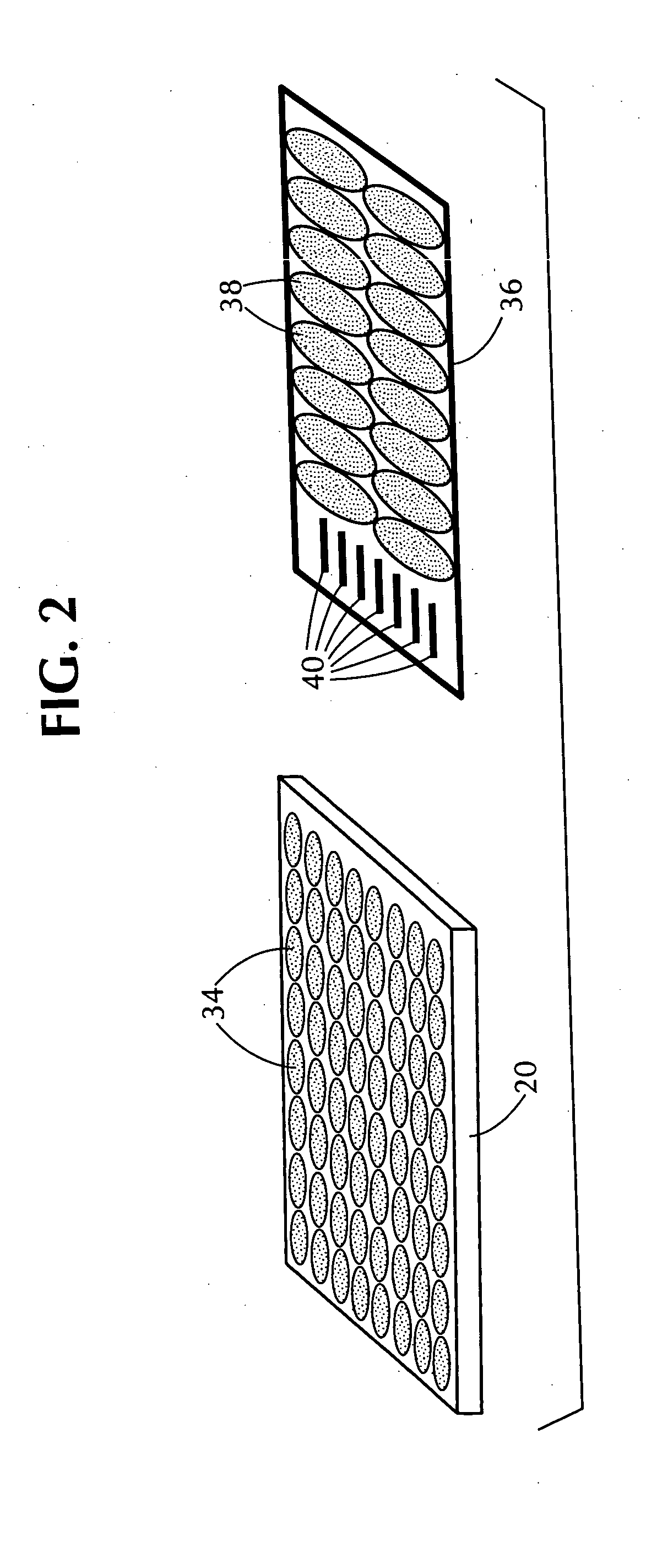
[0242] In the first set of experiments, independent duplicate experiments were run for each of both negative and positive control stimuli. DMSO, which at 1% concentration is known not to be aneugenic or clastogenic (and thus is a negative control), was incubated with Chinese hamster ovary cells in two different microwells, one on each of two different microwell plates, and further processed under substantially identical conditions (incubation temperature, time, and culture medium, fixing and washing protocols, etc.).
[0243] Using the protocol discussed above, one of the wells in a microwell plate was inoculated with 2,500 Chinese hamster ovary cells in a growth medium containing Cytochalasin B, incubated with 50 ng / ml (nanograms / milliliter) of mitomycin C (as the chemical stimulus) for 24 hours at 37 degrees Centigrade, washed, fixed, permeabilized, sequentially treated with Hoechst 33342 and acridine orange, and washed. The microwell plate containing this well was then read by the ...
example 2
[0248] In the second set of experiments, Chinese hamster ovary cells were used and the only difference in treatment and image acquisition protocol between runs in this set was the stimulus (i.e., chemical agent) with which the cells were incubated. Cytochalasin B was not used is this set of experiments (and, thus, the cells remained virtually all mononucleated throughout the experiment). A single run was made using DMSO and two identical but independent runs were made for each of four other compounds, mitomycin C, Compound A, Compound B, and Compound C.
[0249] Table II, below, shows the resulting data, which illustrate the excellent agreement between determinations made using the process of this invention (i.e., micronuclei frequency determined using this invention, referred to as “Auto MN %”) and manual scoring (micronuclei frequency determined using the “Gold Standard,” referred to as “Manual MN %”). The relative difference, calculated as the absolute value of 100 times (Auto MN %...
PUM
 Abstract
Description
Claims
Application Information