
Remedies for heart diseases

a heart disease and drug technology, applied in the field of therapeutic or prophylactic agents of cardiac diseases, can solve the problems of serious conditions of symptomatic cardiac disease, ischemic myocardial dysfunction, and little known about the mechanism behind apoptosis in myocardial cells, and achieve the effects of reducing cell volume, and suppressing apoptosis cell death

Inactive Publication Date: 2005-01-06
CHUGAI PHARMA CO LTD
View PDF3 Cites 1 Cited by
  • Summary
  • Abstract
  • Description
  • Claims
  • Application Information

AI Technical Summary

Benefits of technology

[0033] Thus, the second objective of the present invention is attained by providing a method for screening the above-described low-molecular weight compounds, antibodies, antisense compounds and oligonucleotides to see if they have the Cl− channel inhibiting effect of the invention and can suppress apoptotic cell death in cardiovascular cells.
[0034] The screening method of the invention is characterized by comprising the steps of inducing apoptosis in cardiovascular cells, treating the cells with a Cl− channel blocker under test, and evaluating the therapeutic and / or prophylactic effect of the blocker under test on cardiac disease by checking to see if it can suppress apoptotic cell death in cardiovascular cells.
[0035] In one embodiment of the present invention, apoptosis is induced in cardiovascular cells and simultaneously with or subsequent to this induction of apoptosis, said cardiovascular cells are treated with a compound under test. Check is made to see if apoptotic cell death is suppressed in the cardiovascular cells in the presence of the compound under test as compared with the case where no such compound is present. In this way, one can screen to see if the compound under test proves the therapeutic and / or prophylactic effect on cardiac disease by suppressing apoptotic cell death in cardiovascular cells.
[0036] Examples of the cardiovascular cells contemplated in the invention include myocardial cells, vascular endothelial cells, vascular smooth muscle cells, fibroblasts, myofibroblasts, pericytes and vascular endothelial progenitor cells, with myocardial cells and vascular endothelial cells being preferred. These can be used either as primary cultures of cells or as cells derived from cell lines. If primary cultures of cells are used as cardiovascular cells, heart and / or blood vessels are taken from animals such as rat, mouse, guinea pig, rabbit, bovine and horse for which primary culture systems have been established. The sampled heart and / or blood vessels are treated with a proteolytic enzyme such as collagenase or trypsin before they are prepared as cell suspensions of a predetermined cell density. If cells derived from cell lines are used as cardiovascular cells, either one of the following generally applicable cell lines can be employed, i.e., myocardial cell line, vascular endothelial cell line, vascular smooth muscle cell line, fibroblast cell line, myofibroblast cell line, pericyte cell line and vascular endothelial progenitor cell line, with myocardial cell line and vascular endothelial cell line being preferred. In the case under consideration, the cell line to be employed is cultured under suitable conditions, then prepared as a cell suspension of a predetermined cell density.
[0037] Methods for inducing apoptosis in cardiovascular cells in the present invention include, but are not limited to, chemical methods employing staurosporine (STS), tumor necrosis factor (TNF), TNF in combination with cycloheximide (CHX), anti-Fas agonist antibody, anticancer agent, hydrogen peroxide, etc.; biological methods employing viruses; physical methods employing ultraviolet light, radiations, warm heat, etc.; and combinations of these methods. The dose and the time of treatment required to induce apoptotic cell death in the above-listed cardiovascular cells depend on the cell to be treated and the substance employed to induce apoptosis and can be appropriately determined by the skilled artisan. If staurosporine is employed, treatment is done at a concentration of 0.3-3 μM for 10 minutes to 24 hours, preferably for 30 minutes to 8 hours, to induce apoptosis; if a mixture of TNF and CHX is employed, 0.1-1 μg / ml of CHX is added to 2-10 ng / ml of TNFα and treatment with the mixture is done for 10 minutes to 24 hours, preferably for 30 minutes to 8 hours.
[0038] In the screening method of the invention, the compound under test may be added simultaneously with the above-described induction of apoptosis or, alternatively, it may be added before or after apoptogenic stimulation by the above-described induction of apoptosis. In order to verify its efficacy and effective dose, the compound under test is tested at several concentrations. It is generally preferred to perform an experiment with the concentration of the compound under test being varied over a range from about 10 μM to about 10 mM. The time period of cell treatment with the compound under test depends on the cell to be treated and can be appropriately determined by the skilled artisan; generally speaking, 2-24 hour treatment is sufficient to test for the apoptosis suppressing effect of the compound under test.

Problems solved by technology

Most cases of cardiac disease are primarily due to structural or functional damage to the coronary artery which causes imbalance between blood supply from the coronary artery and its demand by the myocardium, eventually leading to acute or chronic ischemic myocardial dysfunction.
Once apoptosis has been triggered off, myocardial cells are incapable of avoiding apoptosis and cell death occurs, leading to serious conditions of symptomatic cardiac disease such as angina pectoris, myocardial infarction and heart failure.
In addition, little has been known about the mechanism behind apoptosis in myocardial cells and which compounds are capable of selectively suppressing apoptosis in myocardial cells.

Method used


Image

  • Remedies for heart diseases
  • Remedies for heart diseases
  • Remedies for heart diseases

Examples


example 1

[0082] The protective effect of SITS (4-acetamido-4′-isothiocyanostilbene) on staurosporine-induced cytotoxicity in cultured myocardial cells

[0083] Primary cultures of myocardial cells from SD rats at day 20 of pregnancy were used after being cultivated in D-MEM supplemented with 10% of fetal bovine serum. The myocardial cells were cultured on 24-well multiwell plates in an amount of 0.5 mL per well so as to give a density of 5×10⁵ cells / mL.

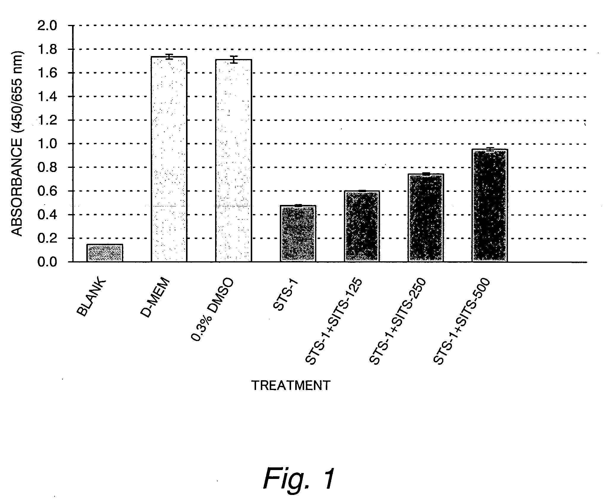
[0084] A control group consisted of three culture media, a cell-free medium (blank), a cell-containing medium alone (D-MEM) and 0.3% DMSO. A test group consisted of two drug-containing culture media, i.e., a solution of 1 μM staurosporine dissolved in 0.3% DMSO (STS-1) and a mixed solution of the 1 μM staurosporine and 125, 250 or 500 μM SITS (STS-1+SITS-125, STS-1+SITS-250, or STS-1+SITS-500). The two groups were cultured in 5% CO2 at 37° C. for 2 hours. Cultivation was continued for another day in D-MEM supplemented with 10% FBS and cell viab...

example 2

[0088] The Protective Effect of SITS on Staurosporine-induced Cytotoxicity in Cultured Myocardial Cells

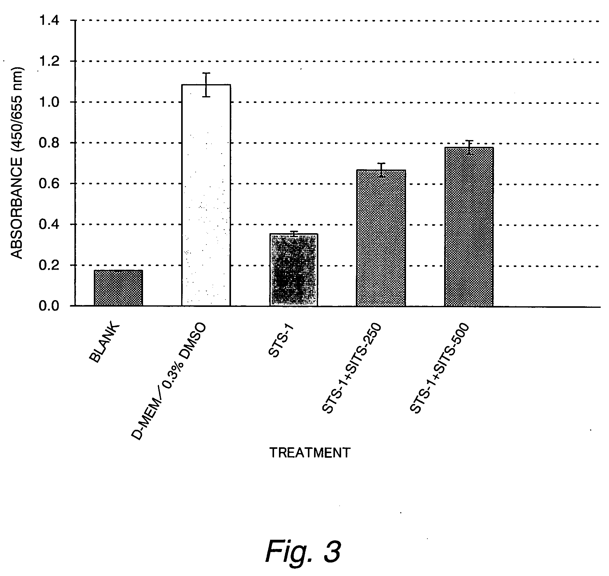
[0089] In this Example, tests were conducted as in Example 1 except that the test group consisted of a 1 μM staurosporine solution (STS-1) and a mixed solution of the 1 μM staurosporine solution and 250 or 500 μM SITS (STS-1+SITS-250 or STS-1+SITS-500) and that the control and test groups were cultured on 96-well multiwell plates rather than the 24-well multiwell plates. The result is shown in FIG. 3.

[0090] As it turned out, it was elucidated that SITS suppresses the drop in cell viability in a dose-dependent manner even when the 96-well multiwell plates were substituted for the 24-well multiwell plates. This shows the possibility of performing high throughput screening of candidate compounds by employing the conditions of Example 2.

example 3

[0091] The protective effect of DIDS (4,4′-diisothiocyanostilbene-2,2′-disulfonic acid) from staurosporine-induced cytotoxicity in cultured myocardial cells

[0092] In this Example, tests were conducted as in Example 1 except that the test group consisted of a 1 μM staurosporine solution (STS-1) and a mixed solution of the 1 μM staurosporine and 62.5, 125 or 250 μM DIDS (STS-1+DIDS-62.5, STS-1+DIDS-125 or STS-1+DIDS-250). The result is shown in FIG. 4.

[0093] As it turned out, it was elucidated that DIDS instead of SITS also suppresses the drop in cell viability in a dose-dependent manner.


PUM

PropertyMeasurementUnit
pHaaaaaaaaaa
volumesaaaaaaaaaa
volumeaaaaaaaaaa

Abstract

This invention aims at providing a method of searching for substances that can selectively suppress apoptosis in cardiovascular cells. This objective is attained by providing a method of screening for therapeutic and / or prophylactic agents of cardiac disease, which comprises the steps of inducing apoptosis in cultured myocardial cells and / or cultured vascular endothelial cells; treating the cells with a Cl− channel blocker under test; and evaluating the therapeutic and / or prophylactic effect of the blocker under test on cardiac disease by checking to see if it can suppress apoptotic cell death in the cardiovascular cells or vascular endothelial cells.

Description

TECHNICAL FIELD [0001] This invention relates to therapeutic or prophylactic agents of cardiac disease. More specifically, the invention relates to therapeutic or prophylactic agents of cardiac disease containing the volume-sensitive outwardly rectifying Cl− channel blockers. [0002] The invention also relates to a method of screening for such therapeutic or prophylactic agents of cardiac disease. More specifically, the invention relates to a method of screening for suitable therapeutic or prophylactic agents of cardiac disease by checking to see if apotosis induced by a known method can be inhibited by a Cl− channel blocker under test. BACKGROUND ART [0003] Most cases of cardiac disease are primarily due to structural or functional damage to the coronary artery which causes imbalance between blood supply from the coronary artery and its demand by the myocardium, eventually leading to acute or chronic ischemic myocardial dysfunction. If myocardial cells become ischemic, it is believe...

Claims


Application Information

Patent Timeline
Patent Type & Authority Applications(United States)
IPC IPC(8): A61K31/00G01N33/50G01N33/68
CPCA61K31/00G01N33/5008G01N33/5061G01N2510/00G01N33/6872G01N2500/10G01N33/5064A61P9/00A61P9/04A61P9/10A61P43/00
Inventor OKADA, YASUNOBUTANABE, SHIGERU
Owner CHUGAI PHARMA CO LTD
Learn More