
Phosphatase associated with metastasis

a phosphatase and metastasis technology, applied in the field of cancer diagnostics and therapeutics, can solve the problems that no gene has been consistently and specifically activated in liver metastases, and achieves the effect of improving the sensitivity and safety of patients

Active Publication Date: 2005-03-03
THE JOHN HOPKINS UNIV SCHOOL OF MEDICINE
View PDF7 Cites 25 Cited by
  • Summary
  • Abstract
  • Description
  • Claims
  • Application Information

AI Technical Summary

Benefits of technology

[0008] According to another embodiment of the invention a method is provided to aid in the prediction of metastasis. Amplification of a first chromosomal segment encoding protein tyrosine phosphatase type IVA, member 3, is detected in a tissue sample of a human. A hybridization probe which hybridizes to all or a portion of a gene encoding protein tyrosine phosphatase type IVA member 3 or primers which amplify all or a portion of protein tyrosine phosphatase type IVA is used to detect the amplification. Detection of such amplification suggests propensity of the tissue to metastasize.
[0010] Also provided by the present invention is an embodiment which can be used as an aid in the prediction of metastasis. Excess expression of a gene is detected in a stool specimen of a human patient having a gastrointestinal neoplasm. The gene is selected from the group consisting of protein tyrosine phosphatase type IVA member 3, FLJ23603, LOC54675, ZD52F10, DNAJ domain-containing, GRO3 oncogene / T45117 hU1-70K protein, attractin, Bc1-2 binding component 3, nuclear receptor subfamily 4, mitogen activated protein kinase 8 interacting protein 2, hairy (Drosophila)-homolog, LUC7 (S. cerevesiae)-like, transducin-like enhancer of split 2, homolog of Drosophila E (sp1), adipose differentiation-related protein, keratin 17, casein kinase 2, alpha prime polypeptide, minichromosome maintenance deficient (S. cerevisiae) 7, v-jun avian sarcoma virus 17 oncogene homolog / LSFR2 gene 2 / MGC2550 protein, plexin B1, transforming growth factor, beta 1ESTs, similar to GTP-rho binding protein 1 (rhophilin), (Drosophila)-like homeo box 1mago-nashi (Drosophila) homolog, proliferation-associated, putative Rab5-interacting protein vascular endothelial growth factor, PTD008 protein, protein / ribosomal protein L10, weel+ (S. pombe) homolog / protein×013, cDNA: FLJ12683, PTK7 protein tyrosine kinase 7v-fos FBJ murine osteosarcoma viral oncogene homolog B, FLJ20297 protein SET translocation (myeloid leukemia-associated), chaperonin containing TCP1, subunit 6A (zeta 1), ataxin 2 related protein, cyclin-dependent kinase inhibitor 3 (CDK2-associated dual specificity phosphatase), and matrix metalloproteinase 14 (membrane-inserted). Excess expression is elevated expression in the stool specimen relative to a stool specimen of a human who does not have a gastrointestinal neoplasm. Excess expression permits the identification of the human patient as having a metastasis or a propensity for a cancer to metastasize.

Problems solved by technology

However, no gene has been shown to be consistently and specifically activated in liver metastases, the lesions that are usually responsible for the death of colorectal cancer patients.

Method used


Image

  • Phosphatase associated with metastasis
  • Phosphatase associated with metastasis
  • Phosphatase associated with metastasis

Examples


example 1

SAGE Analysis of Liver Metastases

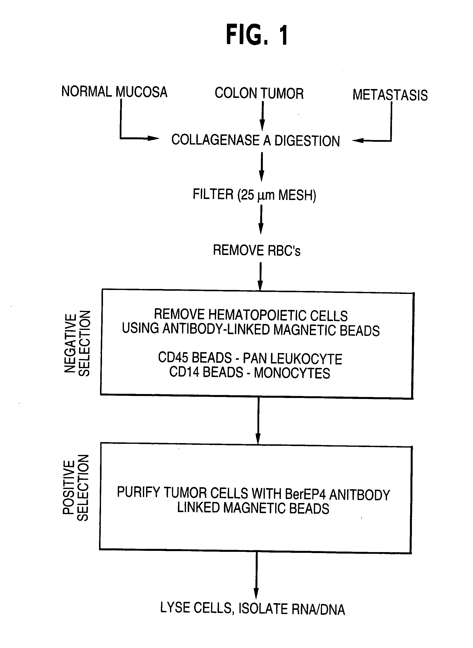
[0033] To learn which genes might be involved in this process, we performed global gene expression profiles of liver metastases using Serial Analysis of Gene Expression (SAGE) technology (5). We first prepared a SAGE library from microdissected metastases (6). Surprisingly, we found that many of the transcripts identified in these libraries were characteristic of normal hepatic or inflammatory cells, precluding quantitative analysis (7). To produce a more specific profile of metastatic epithelial cells, we developed an immunoaffinity fractionation procedure to purify colorectal epithelial cells from contaminating stromal and hepatic cells (8). A SAGE library was prepared from cells purified in this manner, yielding ˜95,000 tags representing at least 17,324 transcripts (6). These tags were compared to ˜4 million tags derived from diverse SAGE libraries, particularly those from normal and malignant (but non-metastatic) colorectal epithelium (9). One h...

example 2

Real-time PCR on Multiple Metastatic Lesions

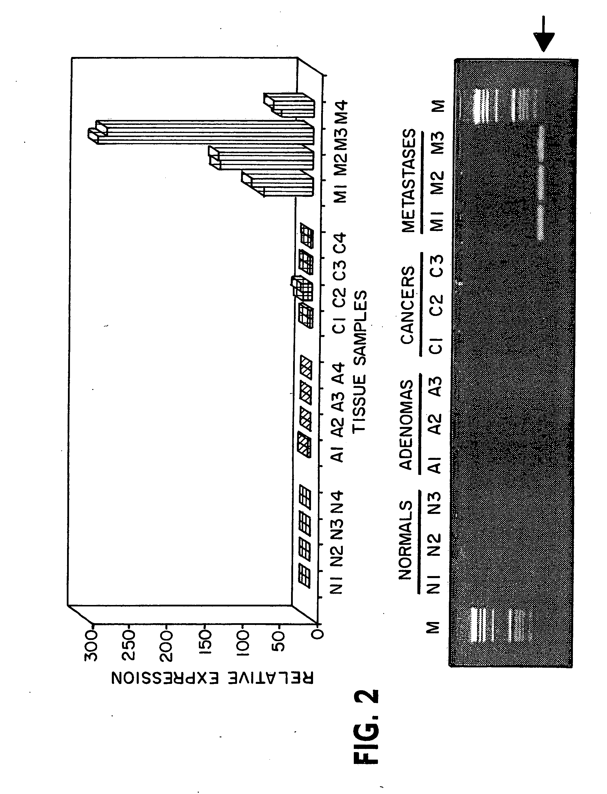
[0038] Transcripts that were enriched rather than depleted in the metastasis library have obvious diagnostic and therapeutic potential. We therefore selected 38 of the most interesting enriched transcripts for further analysis (Table 1) (11). To confirm the SAGE data, we compared the expression of these transcripts in several microdissected metastases, primary cancers, pre-malignant adenomas, and normal epithelium by quantitative, real-time PCR (8). Though all 38 of these transcripts were found to be elevated in at least a subset of the metastatic lesions tested, only one gene, PRL-3, was found to be consistently overexpressed.

[0039] In Table 1, SAGE TAG refers to the 10 bp sequence immediately adjacent to the NlaIII restriction site (CATG) in each transcript. SAGE libraries were constructed from two normal colonic tissues (one library containing 48,479 tags, the other 49,610 tags), two primary colorectal cancers (one library containing ...

example 3

Correlation of Expression of PRL-3 with Metastasis

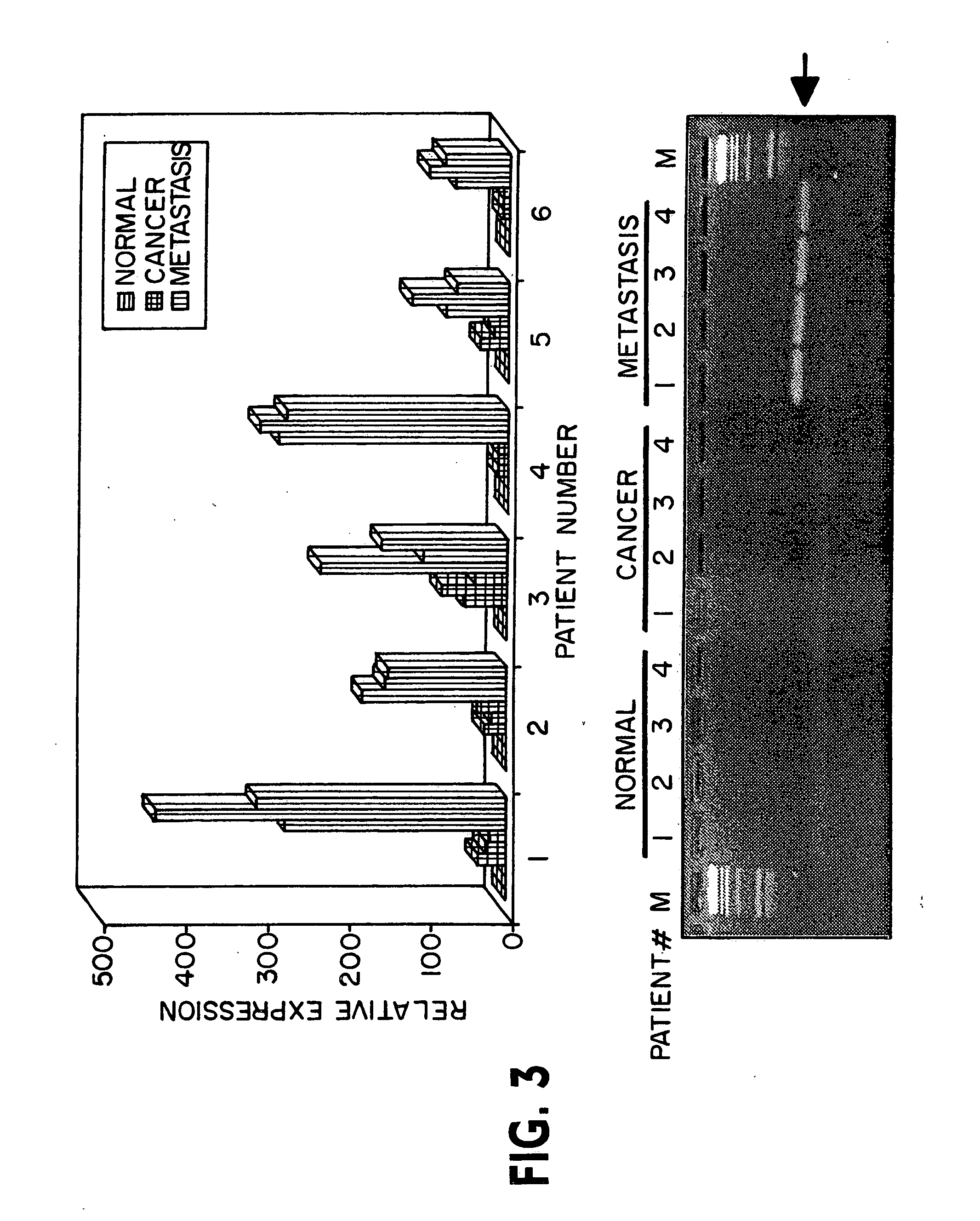
[0040] PRL-3 (also known as PTP4A3) encodes a small 22 kD tyrosine phosphatase that is located at the cytoplasmic membrane when prenylated at its carboxyl terminus and in the nucleus when it is not conjugated to this lipid (12). Among normal human adult tissues, it is expressed predominantly in muscle and heart (13). Although PRL-3 had not been linked previously to human cancer, overexpression of PRL-3 has been found to enhance growth of human embryonic kidney fibroblasts (13) and overexpression of PRL-1 or PRL-2, close relatives of PRL-3, has been found to transform mouse fibroblasts and hamster pancreatic epithelial cells in culture and promote tumor growth in nude mice (14, 15). Based on this information, our preliminary expression data, and the importance of other phosphatases in cell signaling and neoplasia, we examined PRL-3 in greater detail. We first investigated PRL-3 expression in epithelial cells purified from colorectal ...


PUM

PropertyMeasurementUnit
adhesionaaaaaaaaaa
timeaaaaaaaaaa
fluorescentaaaaaaaaaa

Abstract

Among the genes identified, in a comparison of the global gene expression profile of metastatic colorectal cancer to that of primary cancers, benign colorectal tumors, and normal colorectal epithelium, the PRL-3 protein tyrosine phosphatase gene was of particular interest. It was expressed at high levels in each of 18 cancer metastases studied but at lower levels in non-metastatic tumors and normal colorectal epithelium. In three of twelve metastases examined, multiple copies of the PRL-3 gene were found within a small amplicon located at chromosome 8q24.3. These data suggest that the PRL-3 gene is important for colorectal cancer metastasis and provides a new therapeutic target for these intractable lesions.

Description

[0001] This invention was made with support from NIH grants CA57345 and CA43460. The U.S. government therefore retains certain rights in the invention.BACKGROUND OF THE INVENTION [0002] 1. Field of the Invention [0003] The invention relates to the field of cancer diagnostics and therapeutics. In particular it relates to a gene which appears to be significantly associated with the progression of cancer to more advanced stages, including metastasis. [0004] 2. Background of the Prior Art [0005] Metastasis is the neoplastic process responsible for most deaths from cancer, as the primary tumors can usually be surgically removed. Metastatic cells cytoskeletal changes, loss of adhesion, enhanced motility, and express proteolytic enzymes that degrade the basement membrane (1-3). However, much remains to be learned about this lethal process and further progress is contingent upon identifying novel genes and pathways that are consistently and specifically altered in metastatic lesions. [0006]...

Claims


Application Information

Patent Timeline
Patent Type & Authority Applications(United States)
IPC IPC(8): A61K39/00A61K39/395G01N33/50A61P35/00C12N9/16C12N15/09C12Q1/42C12Q1/68C12Q1/6886G01N33/15G01N33/543G01N33/573G01N33/574
CPCA61K39/00C12N9/16C12Q2600/158C12Q2600/112C12Q2600/136C12Q1/6886A61P35/00
Inventor VOGELSTEIN, BERTKINZLER, KENNETHSAHA, SAURABHBARDELLI, ALBERTO
Owner THE JOHN HOPKINS UNIV SCHOOL OF MEDICINE
Learn More