Multipotent amniotic fetal stem cells

a technology of amniotic fluid and stem cells, which is applied in the field of stem cell research, can solve problems such as tumorigenic attributes

Active Publication Date: 2005-03-10
RGT UNIV OF CALIFORNIA
View PDF15 Cites 256 Cited by
  • Summary
  • Abstract
  • Description
  • Claims
  • Application Information

AI Technical Summary

Problems solved by technology

Unfortunately, researchers have found that non-embryonic types of stem cells (“adult stem cells”) are not as capable of differentiating into many different tissue types as are embryonic stem cells, so embryonic stem cells still have many advantages over the use of adult stem cells.
However, one obstacle with the isolation of embryonic stem cells is that the cells are derived from embryos at the “blastocyst” stage.
The tumorigenic attributes of hES cells are not frequently addressed, though this issue may burden their use in replacement cell therapy in the future.
The political, moral and ethical issues around hES cells and their tumorigenic properties, as well as the perceived difficulties of expanding undifferentiated adult stem cells in culture, while maintaining a genetically normal genome, are major barriers in the development of human cell replacement therapy.

Method used


Image

  • Multipotent amniotic fetal stem cells
  • Multipotent amniotic fetal stem cells
  • Multipotent amniotic fetal stem cells

Examples


example 1

Isolation and Expansion of Undifferentiated Cells Derived from Amniotic Fluid

[0056] Approximately 2 to 5 ml of fresh amniotic fluid was harvested from women undergoing routine amniocentesis at 16 to 21 weeks of pregnancy (2nd trimester). Second trimester amniotic fluid contained approximately 1-2×10⁴ live cells per ml. The cells were pelleted in a clinical centrifuge and resuspended in 15 ml “MAFSC” medium. MAFSC medium was composed of low glucose Dulbecco Modified Eagle's Medium (GIBCO, Carlsbad, Calif.) and MCDB 201 medium (SIGMA, Saint Louis, Mo.) at a one to one ratio and contained 2% Defined Fetal Calf Serum (HYCLONE, Logan, Utah), 1× insulin-transferrin-selenium, linoleic-acid-bovine-serum-albumin (ITS+1, SIGMA), 1 nanomolar dexamethasone (Sigma), 100 μm ascorbic acid 2-phosphate (Sigma), 4 μm / ml gentamycin, 10 ng / ml of rhEGF (R&D Systems, Minneapolis, Minn.), 10 ng / ml rrPDGF-BB (R&D) and 10 ng / ml rhFGF-basic (R&D). The wells of 6-well culture dishes were prepared for cell pl...

example 2

Morphological Characterization of Cell Types

[0059] Cultured amniotic fluid-derived cells were tested for cell surface and differentiation markers and were karyotyped. These cells were found to be immortal or near-immortal and were named Multipotent Amniotic Fetal Stem Cells (MAFSC).

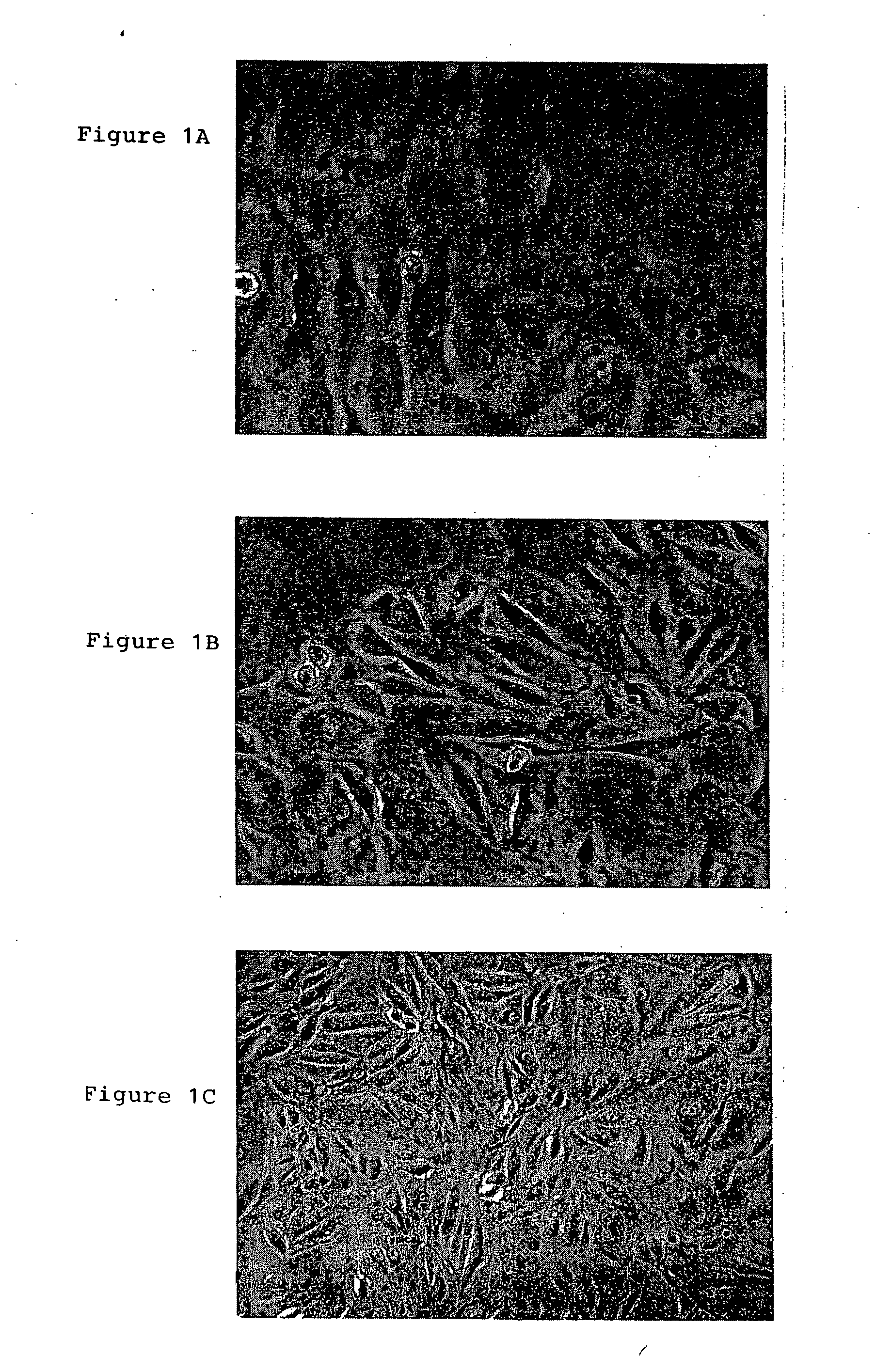
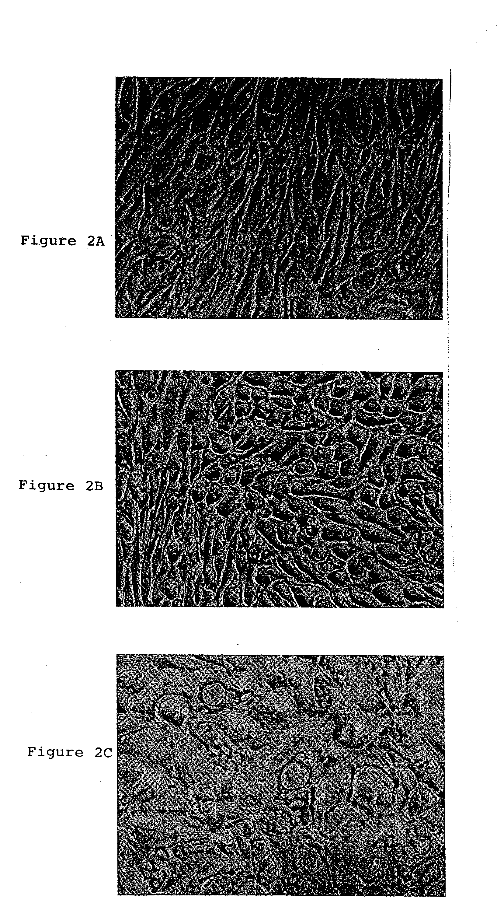
[0060] All of >80 amniotic fluid sample harvests of 5 ml gave rise to at least one adherent MAFSC colony and continuous culture. The majority of sample harvests gave rise to 3-4 individual clones. Among the individual clones, different colonies / cultures had diverse colony morphologies, as shown in FIG. 1. Some cultures had a flat, epihelial morphology (FIG. 1A). Others had a fibroblastic morphology (FIGS. 1B, 1C). Both the epithelioid and the fibroblastic classes of cultures senesced after ˜60 population doublings (PD), yielding a maximum of 1018 cells, unless the cells were immortalized by the expression of the human TERT (telomerase) gene that maintained the length of the cells' telomeres. Indeed, mor...

example 3

FACS Analysis of MAFSC Cells

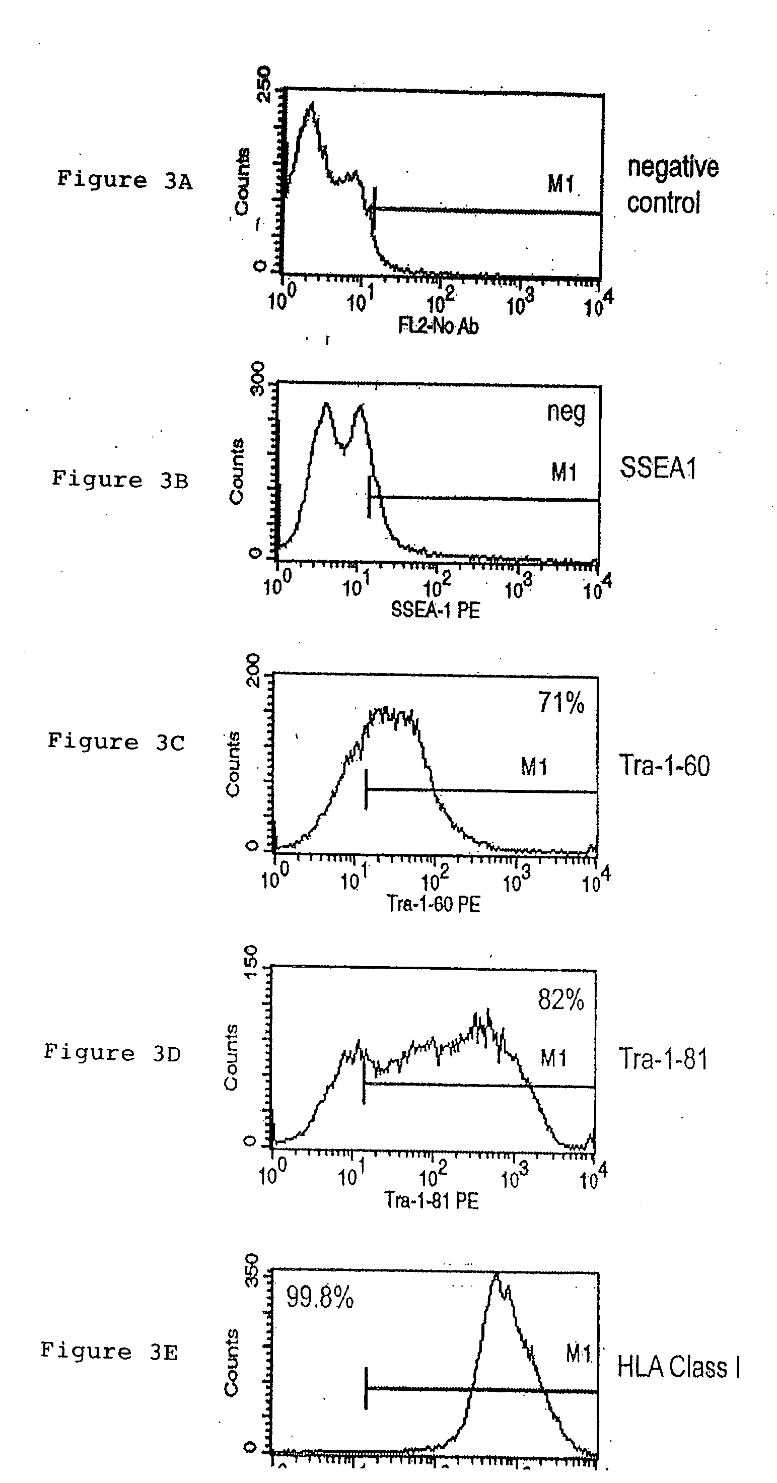
[0062] Cells were prepared for FACS analysis by trypsinizing to remove them from the tissue culture flask, washing in buffer, HBSS, 2% BSA, 0.1% sodium azide, then resuspended in 100 μL of the same buffer. For intracellular antigens (i.e. Oct-4), the cells were fixed and permeablized using Beckman-Coulter IntraPrep reagents, as suggested by the manufacturer. Primary antibodies specific for the indicated cell-surface or intracellular marker were added at a 1:10 dilution and incubated for 30 minutes at room temperature, then washed. For samples using primary antibodies that were not fluorescently-conjugated, the cells were then resuspended in 250 μL of buffer and the appropriate fluorescent-labeled secondary antibody was added at a 1:250 dilution and incubated for 30 minutes at room temperature. Labeled cells were washed and resuspended in buffer or 1% paraformaldehyde for analysis by a FACSCalibur flow cytometer. The data obtained from this analysis were ...


PUM

PropertyMeasurementUnit
doubling timeaaaaaaaaaa
compositionaaaaaaaaaa
surfaceaaaaaaaaaa

Abstract

A source of multipotent amniotic fluid / fetal stem cells (MAFSCs) is disclosed. MAFSC are of fetal origin and have a normal diploid karyotype. These cells are characterized by the following cell surface markers: SSEA3, SSEA4, Tra-1-60, Tra-1-81, Tra-2-54, HLA class I, CD13, CD44, CD49b, CD105 and are distinguished by the absence of the antigen markers CD34, CD45, and HLA Class II, but are distinguished from mouse embryonic stem cells in that these cells do not express the cell surface marker SSEA1. MAFSC express the stem cell transcription factor Oct-4. MAFSC cells can be propagated for an indefinite period of time in continuous culture in an undifferentiated state. The MAFSCs have the ability to differentiate in culture in a regulated manner, into three or more subphenotypes. Cells can then be differentiated into endodermal, mesodermal and ectodermal derived tissues in vitro and in vivo. A method for isolating, identifying, expanding and differentiating MAFSCs is disclosed.

Description

RELATED APPLICATIONS [0001] This application claims priority under 35 U.S.C. §119 to U.S. Provisional Application No. 60 / 495,513, filed Aug. 14, 2003, and U.S. Provisional Application No. 60 / 495,437, filed Aug. 14, 2003 the disclosures of which are incorporated by reference herein in their entireties.BACKGROUND OF THE INVENTION [0002] 1. Field of the Invention [0003] The invention relates to the field of stem cell research, and more particularly to the isolation of stem cells from amniotic fluid. Stem cells derived from amniotic fluid can then be differentiated to many types of cells or tissues. [0004] 2. Description of the Related Art [0005] Stem cells have the ability to divide for indefinite periods in culture and to give rise to specialized cells. Stem cells can give rise to many types of differentiated cells, and may be useful to treat many types of diseases. There are several types of stem cells, such as embryonic stem cells, which are undifferentiated cells from the embryo, a...

Claims


Application Information

Patent Timeline
Patent Type & Authority Applications(United States)
IPC IPC(8): A01N1/02C12NC12N5/073C12N5/074G01N33/48G01N33/50G06F19/00
CPCC12N5/0605C12N5/0607C12N2500/25C12N2501/39C12N2501/115C12N2501/135C12N2501/11
Inventor HAAS, MARTIN
Owner RGT UNIV OF CALIFORNIA
Why Eureka
Try Eureka