
Method and apparatus for detection of microscopic pathogens

Inactive Publication Date: 2005-05-19
WISCONSIN ALUMNI RES FOUND
View PDF13 Cites 60 Cited by
  • Summary
  • Abstract
  • Description
  • Claims
  • Application Information

AI Technical Summary

Benefits of technology

[0015] In accordance with the present invention, microscopic pathogens such as bacteria and viruses may be detected in a simple and efficient manner. The detection of a pathogen can be carried out by personnel who have minimal training and without requiring specialized laboratory facilities or equipment. Detection is provided with accurate readout in a manner that is faster than conventional serological tests. It is possible to screen for multiple microscopic pathogens in a single test.
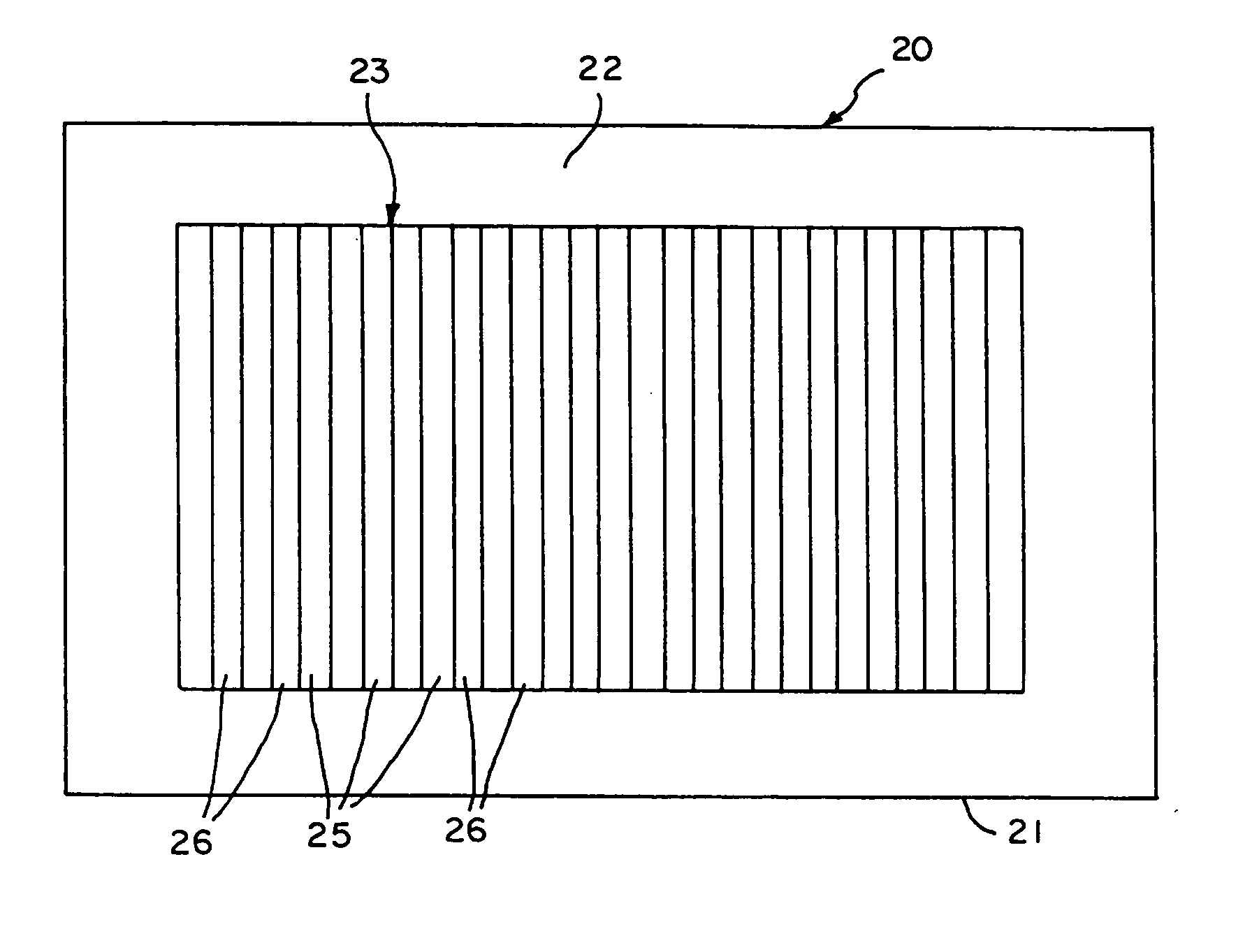
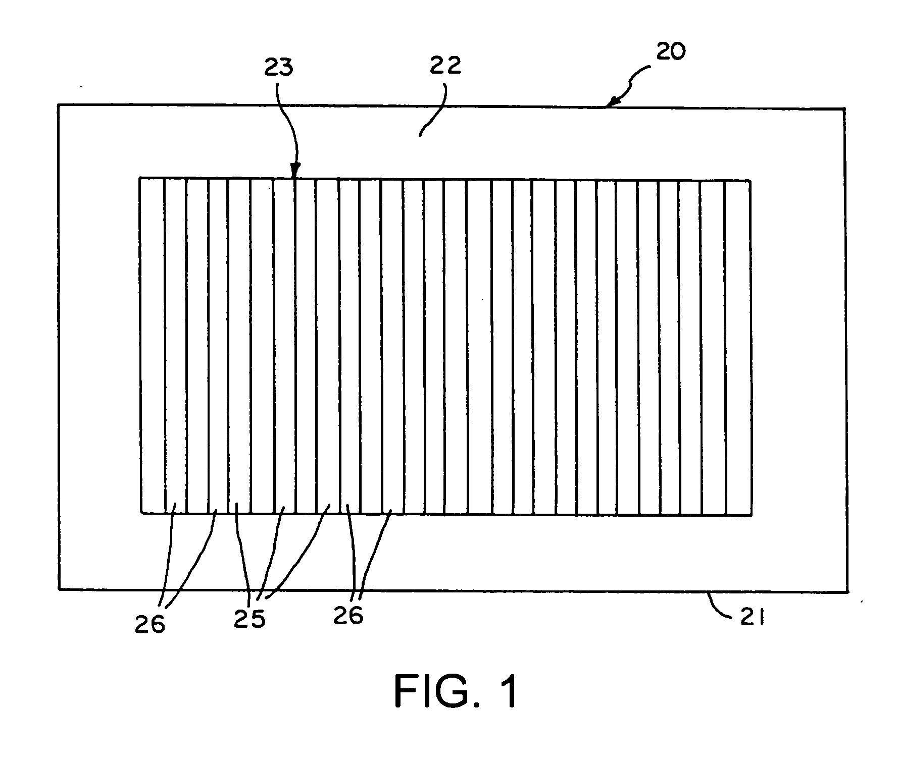
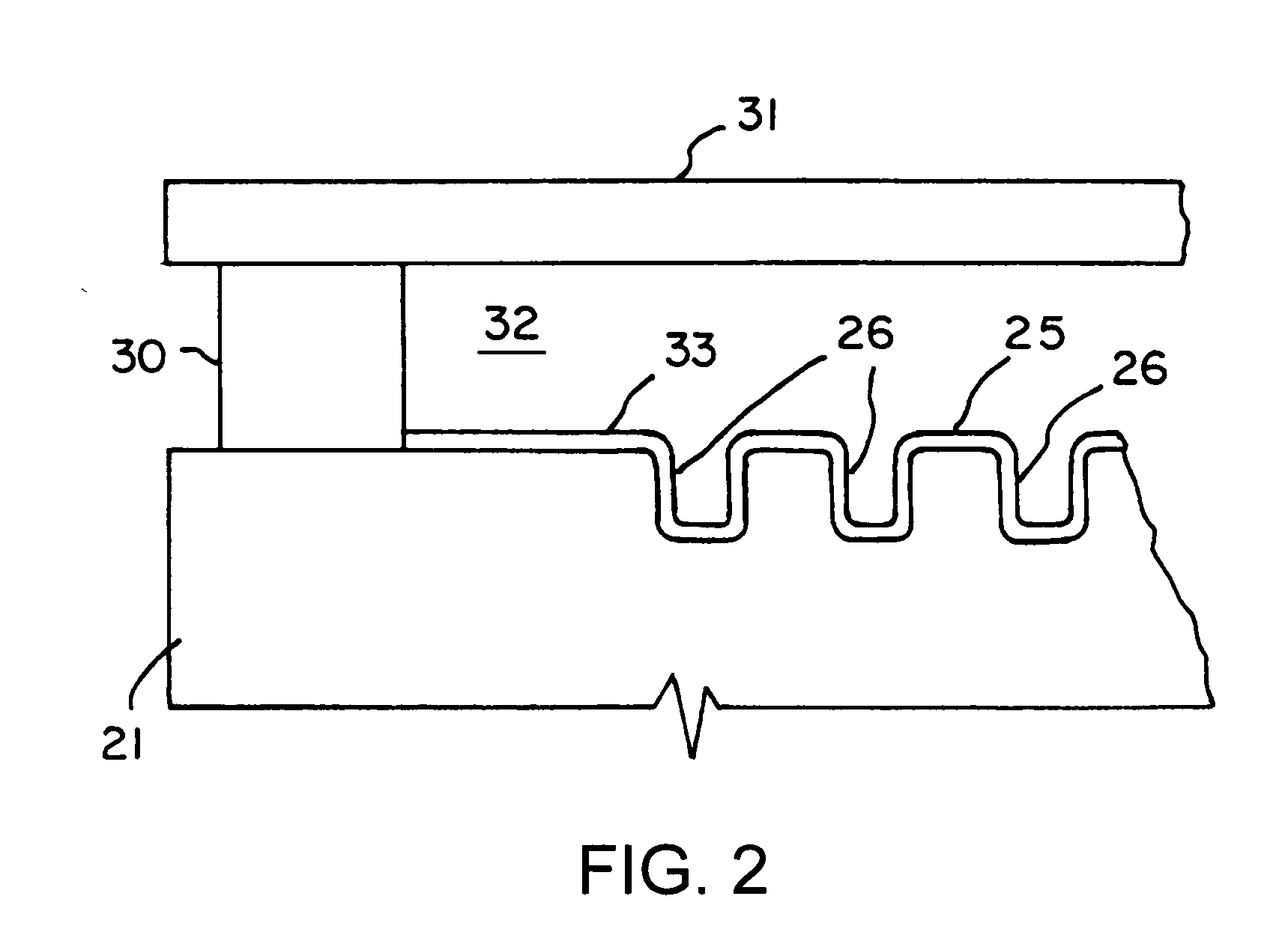
[0018] To test a sample for the presence of the selected pathogen, the sample is contacted to the surface of the detection region to permit the pathogen particles, if present, to be bound to the surface by the binding agents and to occupy the depressions. A liquid crystal material is thereafter applied to the detection region that will be aligned by the microstructures on the surface of the substrate in the absence of binding of particles to the surface. Where no particles are present on the surface, the liquid crystal material is aligned and appears uniform and dark when visually examined, typically by utilizing an appropriate polarizing viewing material. If the specific pathogen is present in the sample, it will be bound to the binding agents and will substantially occupy the depressions on the surface of the substrate. The occupation of the depressions by the particles disrupts the uniform alignment of the liquid crystal material, with the result that the detection region appears relatively brightly colored when viewed with the appropriate polarizing material. In this manner, an observer can readily and easily determine whether or not the specific pathogen is present in the sample. The depressions on the substrate are sufficiently large compared to molecular material such as proteins which may be bound to the surface, and thus the non-specific binding of proteins or other molecular materials does not disrupt the uniform alignment of the liquid crystal material.

Problems solved by technology

The occupation of the depressions by the particles disrupts the uniform alignment of the liquid crystal material, with the result that the detection region appears relatively brightly colored when viewed with the appropriate polarizing material.

Method used


Image

  • Method and apparatus for detection of microscopic pathogens
  • Method and apparatus for detection of microscopic pathogens
  • Method and apparatus for detection of microscopic pathogens

Examples


examples

Materials

[0061] Glass microscope slides used were Fisher's Finest, Premium Grade and were obtained from Fisher Scientific (Pittsburgh, Pa.). Tridecafluoro-1,1,2,2-tetrahydrooctyl trichlorosilane was purchased from Gelest (Tullytown, Pa.). 1-Decanethiol, 1-hexadecanethiol, and mineral oil were obtained from Aldrich (Milwaukee, Wis.). The following were used as thermally- or UV-curable prepolymers: poly(dimethylsiloxane) (PDMS, Sylgard® 184, Dow Corning Co. (Midland, Mich.)); epoxy resin (2-Ton® Clear Epoxy, Devcon (Danvers, Mass.)); polyurethane (PU, NOA61, Norland Products Inc. (New Brunswick, N.J.)); polycyanoacrylate (PC, J-91, Summers Optical (Fort Washington, Pa.)); and polystyrene (PS, UV-74, Summers Optical (Fort Washington, Pa.)). Bovine serum albumin (BSA, IgG free, lyophilized powder) was obtained from Sigma (St. Louis, Mo.) and used as received. The nematic liquid crystal of 4-cyano-4′-pentylbiphenyl (5CB), manufactured by BDH, was purchased from EM industries (Hawthorne,...


PUM

PropertyMeasurementUnit
Widthaaaaaaaaaa
Widthaaaaaaaaaa
Widthaaaaaaaaaa

Abstract

Detection apparatus for use in the detection of the presence of a selected pathogen in a sample are disclosed. Such apparatus include: a substrate with a detection region on a surface thereof, the detection region having microstructures including grooves formed therein that will align liquid crystal material in contact therewith, the width and depth of the grooves being in the range of 10 μm or less; a blocking layer on the surface of the detection region of the substrate that does not disrupt the alignment of liquid crystal material in contact therewith, the blocking layer blocking nonspecific adsorption of pathogens to the surface; and a binding agent on the surface of the detection region of the substrate, the binding agent specifically binding the selected pathogen.

Description

CROSS-REFERENCES TO RELATED APPLICATIONS [0001] This application is a divisional of and claim priority to U.S. Ser. No. 09 / 784,232, filed Feb. 15, 2001, pending, which claims priority to U.S. Provisional Application No. 60 / 182,941, filed Feb. 16, 2000, the entire disclosures both of which are incorporated herein by reference in their entireties and for all purposes as if fully set forth herein.GOVERNMENT RIGHTS [0002] This invention was made with United States government support awarded by the following agency: ONR N00014-97-0702. The United States has certain rights in this invention.FIELD OF THE INVENTION [0003] This invention pertains generally to assay devices and methods for the detection of microscopic pathogens such as viruses or bacteria in a sample. BACKGROUND OF THE INVENTION [0004] Methods for detecting the presence of biological substances and chemical compounds in samples has been an area of continuous development in the field of analytical chemistry and biochemistry. V...

Claims


Application Information

Patent Timeline
IPC IPC(8): G01N33/53G01N33/543G01N33/545G01N33/552G01N33/553G01N33/566G01N33/569
CPCB82Y30/00G01N33/54373Y10S436/807G01N33/56911G01N33/569
Inventor ABBOTT, NICHOLAS L.SKAIFE, JUSTIN J.
Owner WISCONSIN ALUMNI RES FOUND
Learn More