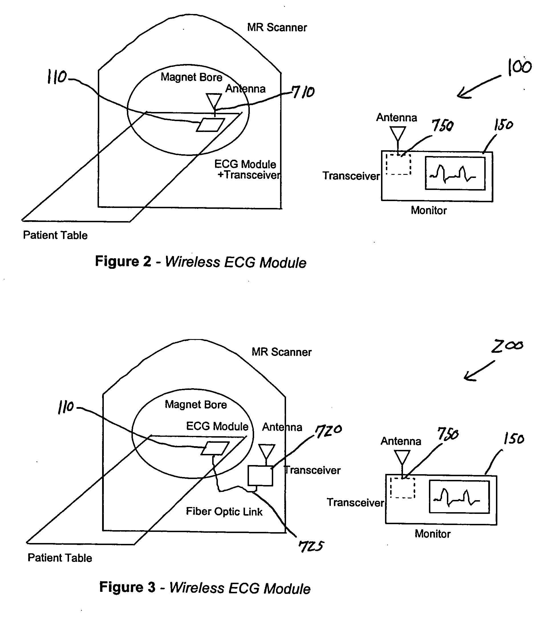
[0025] In a presently preferred embodiment, the invention provides a system for wirelessly communicating physiologic data indicative of the condition of a patient exposed to a scanner of an MR system. The system includes a sensor mechanism, a first
transducer circuit, a first RF transceiver circuit, a second RF transceiver circuit, and a second
transducer circuit. The sensor mechanism is used to acquire the physiologic data from the patient. The first
transducer circuit connects to the sensor mechanism for converting the physiologic data received therefrom from optical format to electrical format. The first RF transceiver circuit is connected to the first transducer circuit for transmitting the physiologic data received therefrom. The second RF transceiver circuit, which is remote from the first RF transceiver circuit, is used to receive the physiologic data transmitted by the first RF transceiver circuit. The second transducer circuit is connected to the second RF transceiver circuit for converting the physiologic data received therefrom from electrical format to optical format, and for conveying the physiologic data to an apparatus remote from the sensor mechanism. The communication between the sensor mechanism and the apparatus via the first and second RF transceiver circuits is accomplished without adversely affecting, or being adversely affected by, operation of the MR system.
[0026] In a related embodiment, the invention provides a system for wirelessly communicating data in an electromagnetically noisy environment. The system includes a first transducer circuit, a first RF transceiver circuit, a second RF transceiver circuit, and a second transducer circuit. The first transducer circuit connects to a first device of a bifurcated system for converting the data received therefrom from optical format to electrical format. The first RF transceiver circuit is connected to the first transducer circuit for transmitting the data received therefrom. The second RF transceiver circuit, which is remote from the first RF transceiver circuit, is used to receive the data transmitted by the first RF transceiver circuit. The second transducer circuit is connected to the second RF transceiver circuit for converting the data received therefrom from electrical format to optical format, and for conveying the data to a second device of the bifurcated system. The scheme of communication employed by the first and second RF transceivers enables the first and second devices to communicate without being adversely affected by noise in the environment.
[0028] In a different embodiment, the invention provides a system for wirelessly communicating data obtained from a sensor module attached to a patient situated within an imaging scanner. The system includes a first transceiver and a second transceiver. The first transceiver is linked to the sensor module for transmitting the data received therefrom. The second transceiver, which is connected to an apparatus remote from the first transceiver, is used to convey to the apparatus the data received from the first transceiver. The first and second transceivers enable the sensor module and the apparatus to communicate without being adversely affected by, or adversely affecting, the operation of the imaging scanner.
[0031] In a presently preferred embodiment, the invention provides a communications module for wirelessly communicating electrocardiographic (ECG) signals obtained from a patient situated in a noisy environment. The module includes at least one RF filter, a lead select network, a
differential amplifier, an
amplifier circuit, a
signal processing circuit, a modulator circuit, a
transmitter circuit, and a filter circuit. The RF filter(s) is linked to a sensor of bioelectric signals for removing therefrom frequencies outside those carrying the bioelectric signals. The lead select network is used to select, in response to control signals, the appropriate lead(s) of a multiple-lead lead-set from which to
pickup selected one(s) of the bioelectric signals. The
differential amplifier is used to derive the ECG signals from the bioelectric signals selected via the network. The
amplifier circuit is used to amplify the ECG signals received from the
differential amplifier, and the
signal processing circuit is used to improve the condition of the ECG signals received from the
amplifier circuit. The modulator circuit digitally modulates a
carrier signal in accordance with the ECG signals it receives from the
signal processing circuit to form a modulated signal therewith. The
transmitter circuit is connected to the modulator circuit for transmitting the modulated signal received therefrom. Connected to the
transmitter circuit, the filter circuit allows the modulated signal to pass while effectively attenuating unwanted frequencies.
[0032] In a related embodiment, the invention also provides a communications module for wirelessly communicating physiologic signals obtained from a patient situated in a noisy environment. The module includes an input conditioning circuit, a
signal processing circuit, a
converter circuit, a transmitter circuit, and a filter circuit. The input conditioning circuit, which links to a sensor of the physiologic signals, is used to adapt the physiologic signals received therefrom for use in the module. The signal
processing circuit improves the condition of the physiologic signals received from the input conditioning circuit, and the
converter circuit converts the physiologic signals received from the signal
processing circuit to digital signals corresponding thereto. The transmitter circuit is connected to the
converter circuit and is used to transmit the digital signals received therefrom. The filter circuit is connected to the transmitter circuit for passing the digital signals and effectively attenuating unwanted frequencies.

