
Single cell assessment of viral infection/replication

a single cell, viral technology, applied in the field of immunology and molecular pharmacology, can solve the problems of no cure for aids, no cure for infections and diseases, and damage to the body's ability to fight infections and diseases, and achieve the effect of high throughput separation

Inactive Publication Date: 2005-06-02
THE BOARD OF TRUSTEES OF THE LELAND STANFORD JUNIOR UNIV
View PDF2 Cites 2 Cited by
  • Summary
  • Abstract
  • Description
  • Claims
  • Application Information

AI Technical Summary

Benefits of technology

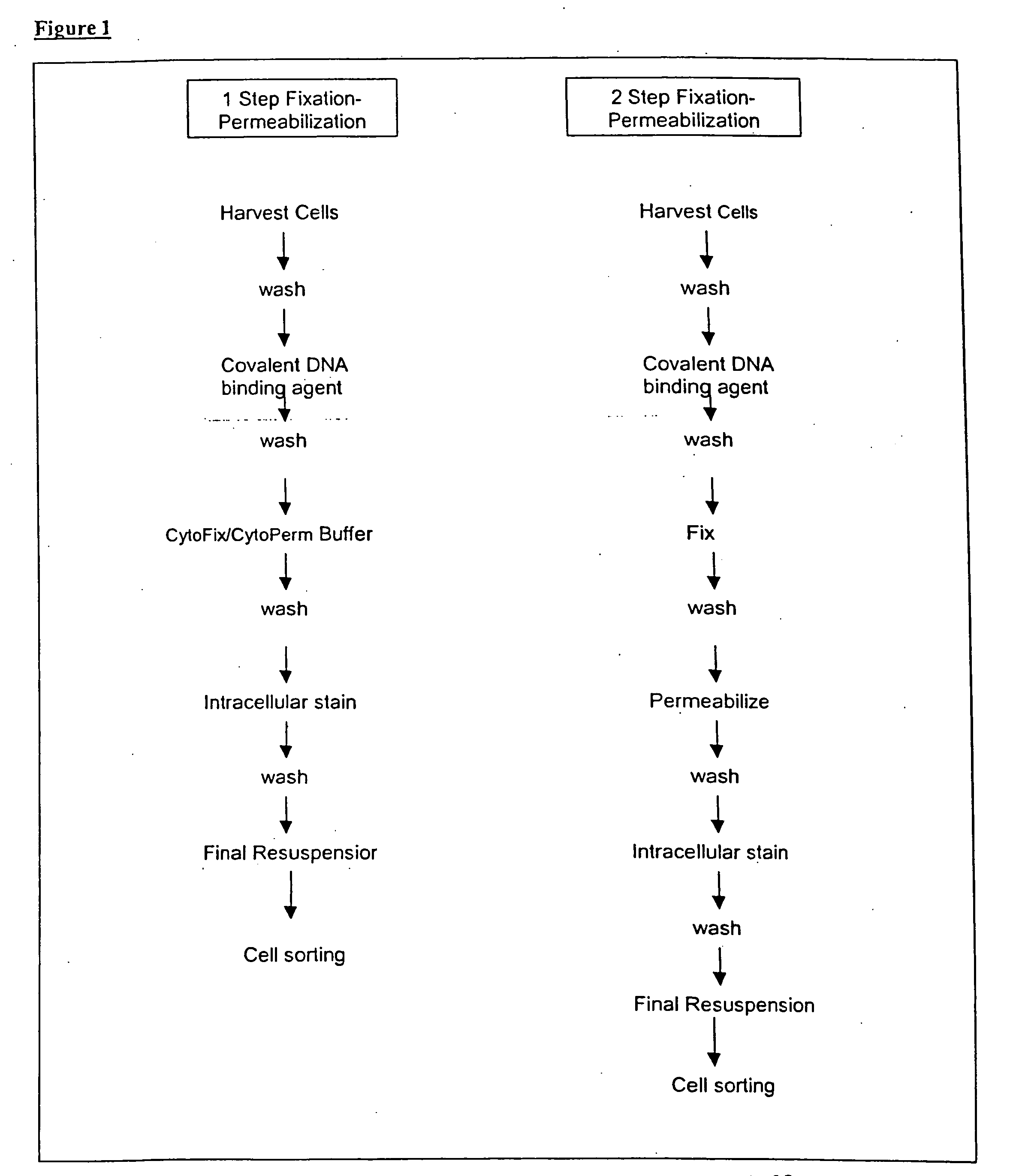
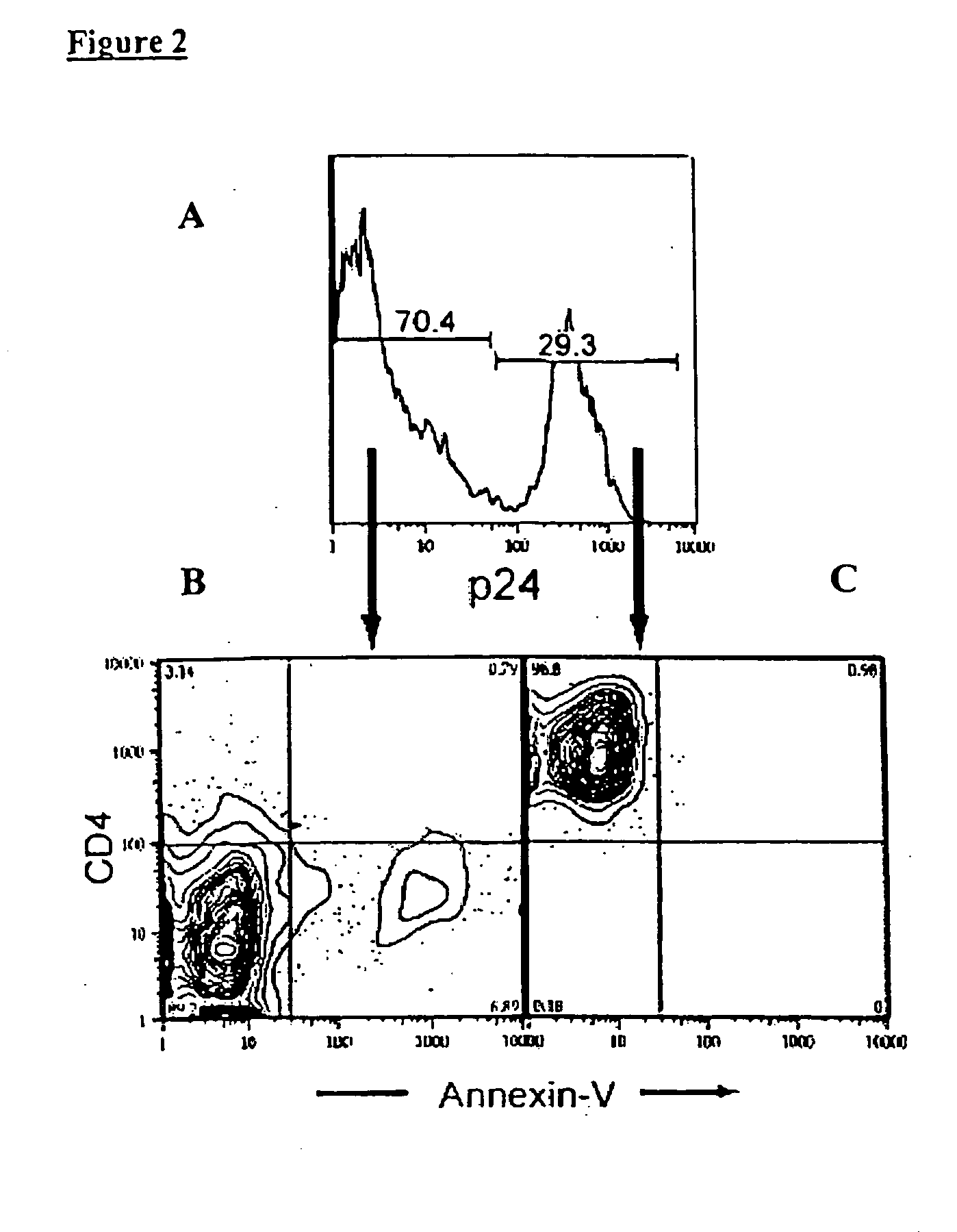
[0012] A principal aspect of the present invention is the design of a technique that allows high throughput separation of virally infected viable cells from dead cells. The method employs antibodies specific for intracellular proteins that are expressed specifically in response to a viral infection, and a nucleic acid binding agent that recognizes preferentially dead cells. The method can be readily adapted for assessing viral infection and / or replication in viable cells, identifying anti-viral agent, and monitoring anti-viral therapy.

Problems solved by technology

By killing or damaging cells of the body's immune system, the virus causes acquired immunodeficiency syndrome (AIDS) and progressively destroys the body's ability to fight infections and disease.
As the virus multiplies and kills immune cells, the body becomes vulnerable to opportunistic infections and other illnesses, ranging from pneumonia to cancer.
There is currently no cure for AIDS.
While the use of bulk measurements and molecular techniques such as RT-PCR and p24 ELISA are useful in detecting intracellular antigens, they cannot quantitatively identify the viable cells harboring viral infection.
However, this technique has not been adapted to effect single cell assessment of viral infection or replication.
This results in false positive readings where non-viable or “dead” cells are identified as infected with an intracellular virus.
Thus, while PI is useful for surface staining alone, it is inadequate for intracellular staining where the permeabilization conditions can cause reversible binding of PI and inadvertently label cells that are not dead.
Therefore, a significant percentage of cells may have died before the cells are processed for further analysis.

Method used


Image

  • Single cell assessment of viral infection/replication
  • Single cell assessment of viral infection/replication
  • Single cell assessment of viral infection/replication

Examples


examples

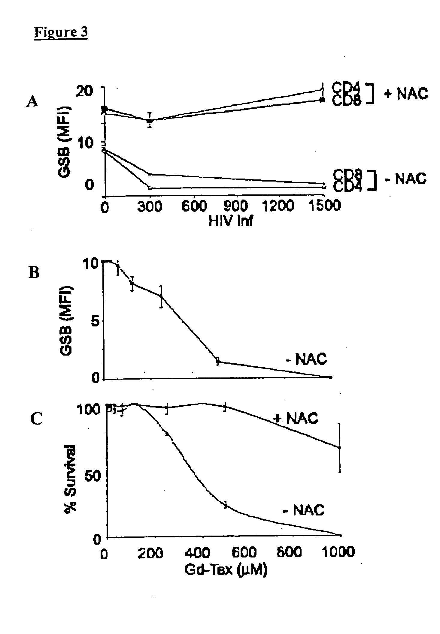
[0084] The methods of the present invention were used to analyze the response of HIV-infected CD4+ cells in IL-2 stimulated cultures in vitro to motexafin gadolinium (Gd-Tex). Gd-Tex is a compound that promotes intracellular oxidative stress and has been reported to localize tumors and to enhance radiation response in animal tumor models.

[0085] Peripheral blood mononuclear cells (PBMC) isolated from healthy donors were first activated in culture with recombinant human IL-2 and infected in vitro by HIV.

[0086] The isolated cells were maintained in complete media (RPMI medium 1640, 10% (vol / vol) FCS, 1% (vol / vol) PSQ) at 37° C. and 5% CO2. Cells were activated with human recombinant IL-2 for 24 hours prior to HIV-1 infection. BSO treatments were preformed at 5 mM for 72 hours and N-acetylcysteine (NAC) treatments were performed at 5 mM for 24 hours. NAC alleviated Gd-Tex toxicity at high Gd-Tex concentrations. The BSO treatment rendered PBMC more sensitive to killing with Gd-Tex. Tre...


PUM


Abstract

This invention relates to methods of separating virally infected viable cells from dead cells using antibodies specific for intracellular proteins and a covalent nucleic acid binding agent. The method can be readily adapted for assessing viral infection and / or replication in viable cells, identifying anti-viral agent, and monitoring anti-viral therapy

Description

CROSS-REFERENCE TO RELATED APPLICATIONS [0001] This application claims priority to two pending U.S. provisional applications Ser. Nos. 60 / 358,425 and 60 / 359,153, filed on Feb. 19, 2002 and Feb. 20, 2002, respectively. These priority applications are hereby incorporated herein by reference in their entirety.STATEMENT OF RIGHTS TO INVENTIONS MADE UNDER FEDERALLY SPONSORED RESEARCH [0002] This invention was made with Government support under contracts awarded by the National Institutes of Health, NIH2R01AI35304. The Government has certain rights in this invention.TECHNICAL FIELD [0003] This invention is in the field of immunology and molecular pharmacology. Specifically, the invention relates to methods of separating virally infected viable cells from dead cells using antibodies specific for intracellular proteins and a covalent nucleic acid binding agent. The method can be readily adapted for assessing viral infection and / or replication in viable cells, identifying anti-viral agent, a...

Claims


Application Information

Patent Timeline
IPC IPC(8): G01N1/30G01N33/569
CPCG01N1/30G01N33/56983G01N33/56988G01N33/56994G01N2333/02G01N2800/52G01N2333/10G01N2333/16G01N2333/18G01N2500/10G01N2333/03Y02A50/30
Inventor PEREZ, OMARNOLAN, GARRY P.
Owner THE BOARD OF TRUSTEES OF THE LELAND STANFORD JUNIOR UNIV
Learn More