
Method and device for minimally invasive implantation of biomaterial

Active Publication Date: 2005-06-16
ETHICON ENDO SURGERY INC
View PDF25 Cites 119 Cited by
  • Summary
  • Abstract
  • Description
  • Claims
  • Application Information

AI Technical Summary

Benefits of technology

[0006] The invention features devices and methods for the delivery of active agents in a subject. The active agent formulation may be stored within an active agent delivery system (e.g., contained in a delivery layer of the system or a reservoir within the controlled active agent delivery system). The active agent formulation may comprise an amount of active agent sufficient for treatment and is stable at body temperatures (e.g., no unacceptable degradation) for the entire pre-selected treatment period. The active agent delivery systems may store the active agent formulation safely (e.g., without dose dumping), provide sufficient protection from bodily processes to prevent unacceptable degradation of the formulation, and release the active agent formulation in a controlled fashion at a therapeutically effective rate to the subject. In use, the active agent delivery system may be implanted in the subject's body at an implantation site, and the active agent formulation may be released from the active agent delivery system to a delivery site within, for example, the peritoneal or abdominal wall. Once released at the delivery site, the active agent formulation may enter the subject through vascularized tissue.
[0007] The present invention may allow a patient greater flexibility of lifestyle, freedom of movement, and the ability to carry out physical activity in a more natural and comfortable manner. The present invention may eliminate the need for frequent injections and the pain associated with multiple daily needle injections, skin irritation, mental anguish suffered by patients having difficulty controlling or maintaining the physiologic parameters pertaining to their clinical condition.
[0008] In one embodiment, the invention comprises an active agent delivery device comprising a tethered delivery device and a minimally invasive method of placing such an active agent delivery device substantially adjacent to vascular tissue. The implantation of such a device may provide a sustained delivery of therapeutic, prophylactic, and / or many other types of materials. Generally, the active agent delivery device may be placed within, for example, the abdominal space, within the omentum, within the peritoneal cavity, a space formed when the parietal and visceral layers of the peritoneum, or between the parietal peritoneum and abdominal wall. Other suitable locations for placement are possible.
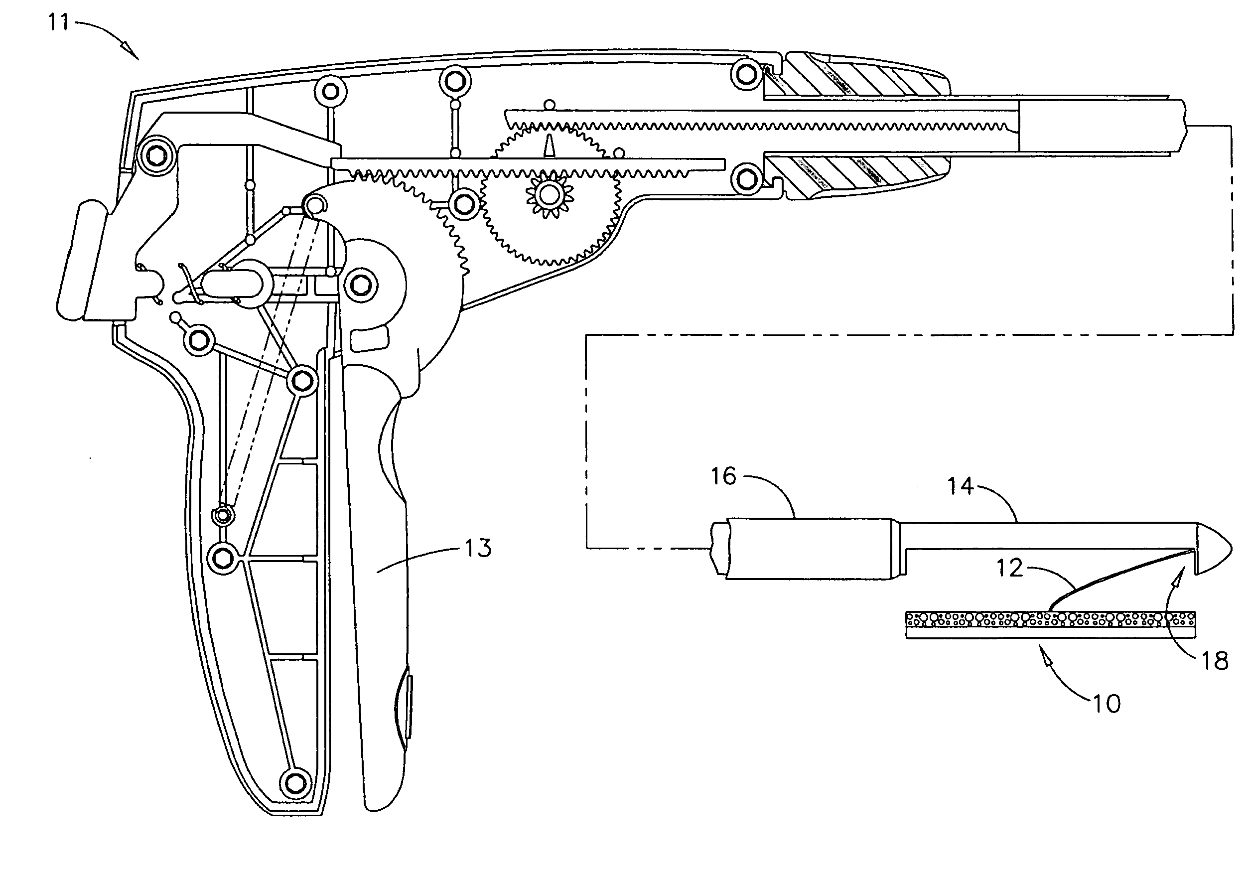
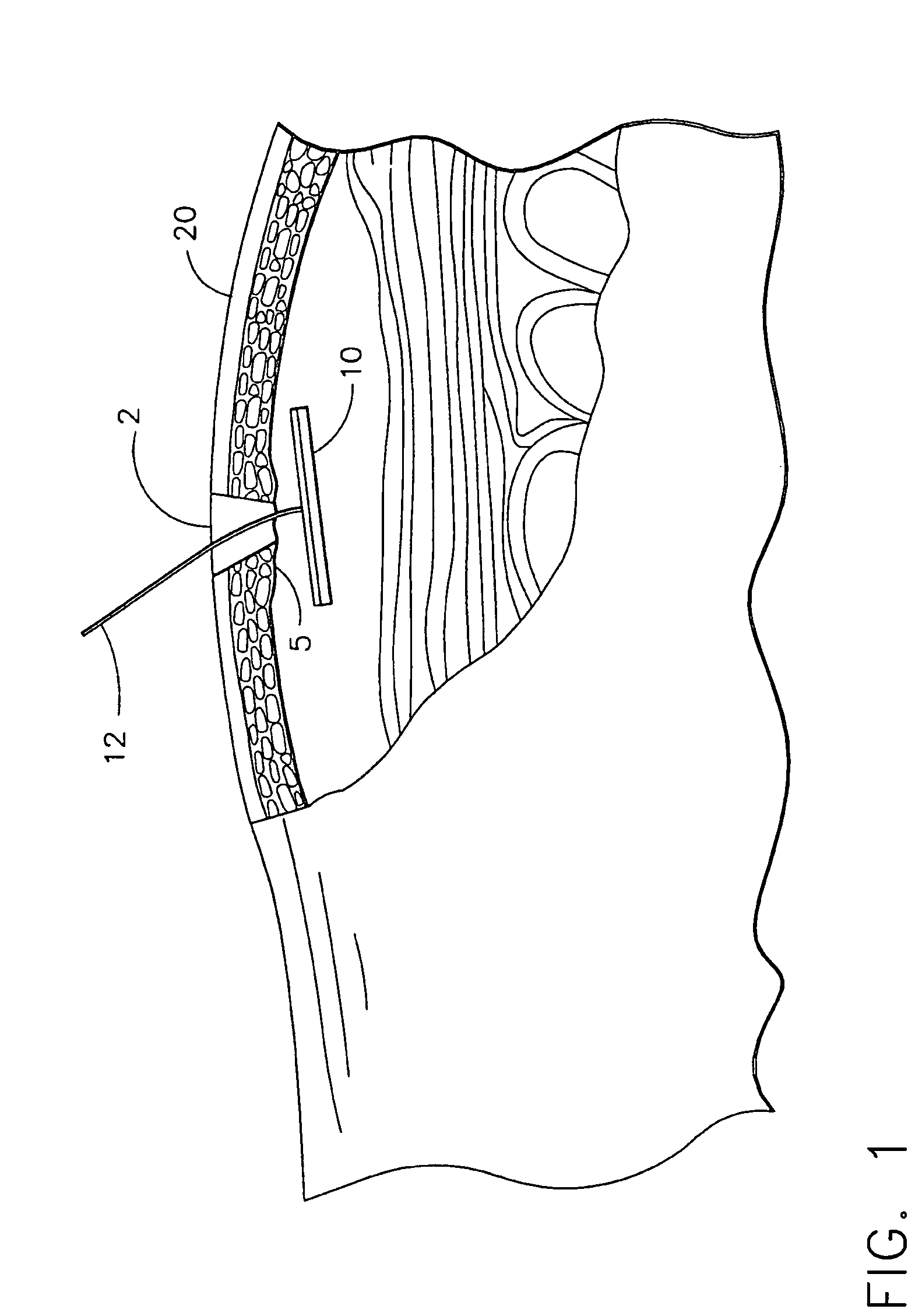
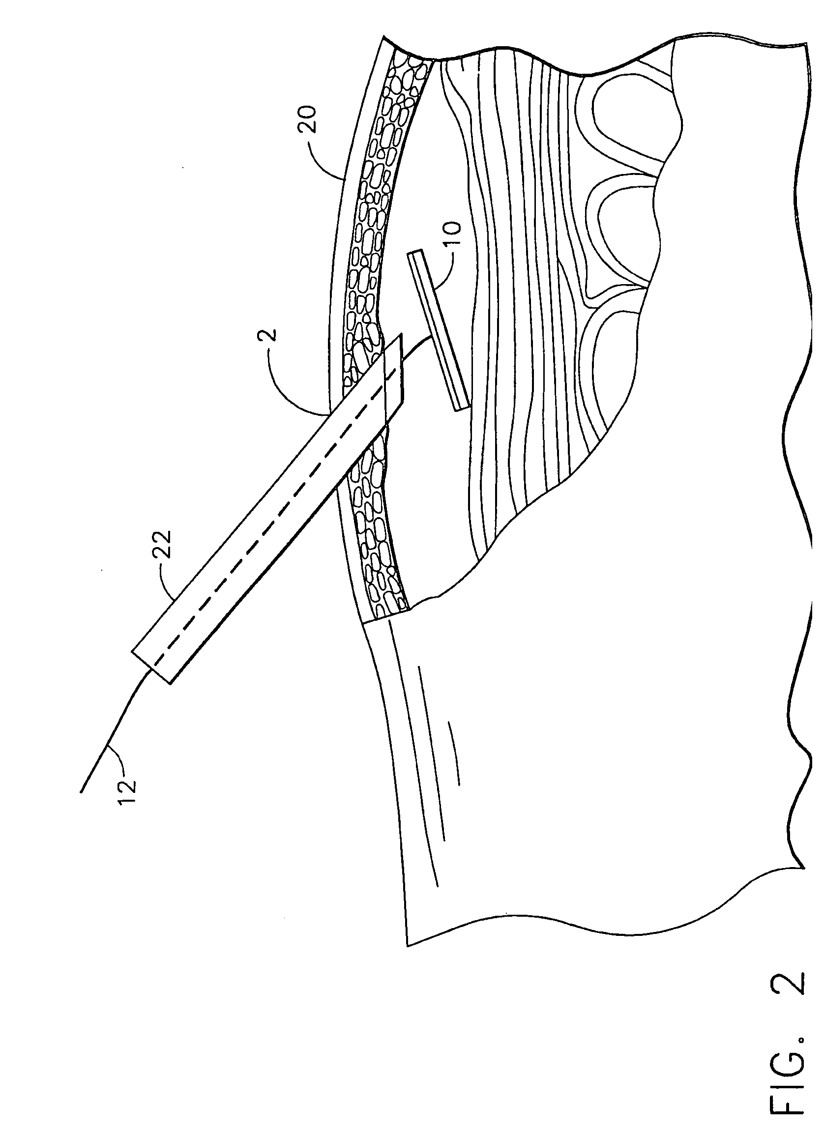
[0010] The active agent delivery device may include a biological construct and a tethering means. The delivery device may be substantially flexible and / or have an adhesive front. The tethering means may be made of a substantially flexible material capable of anchoring the delivery device in substantial planar contact with the vascularized tissue. The delivery device may be percutaneously inserted near vascular tissue such as, for example, peritoneal tissue. When the delivery device has been inserted, the tethering means may be used to pull the delivery device toward the vascular tissue, such that the active agent delivery top surface of the delivery device comes in contact with the vascular tissue. Contact between the active agent delivery surface of the delivery device and the vascular tissue may be maintained by making and keeping a tethering means substantially taut. In an alternate embodiment, an adhesive on the active agent delivery surface of the delivery device capable of substantially adhering to the vascular tissue may be used to keep the delivery device substantially in contact with the vascularized tissue.
[0012] The present inventors have discovered a device that is suitable for the controlled and sustained release of an agent effective in obtaining a desired local or systemic physiological or pharmacological effect. The device includes an implantable active agent delivery system comprising an effective amount of at least one active agent that, once implanted, the device may give a continuous supply of the agent or agents to internal regions of the body without requiring additional invasive penetrations into these regions. Instead, the device may remain in the body and serve as a continuous source of the agent or agents. The device according to the present invention permits prolonged constant release of active agents over a specific period of months (e.g., 1 month, 2 months, 3 months, 6 months) or years (e.g., 1 year, 5 years, 10 years, 20 years) until the agent is substantially used up.

Problems solved by technology

Thus, the art lacks teachings of implantation on or near peritoneal tissue for sustained delivery of materials for non-hernial purposes.
While the art has taught the implantation of cell capsules for sustained delivery of neuroactive substances in the central nervous system, such implantation means are relatively inflexible, and their utility is substantially limited to the central nervous system.
A relatively inflexible implant would not fully take advantage of the vascular nature of tissue such as peritoneal tissue if implanted on or near such tissue, as a relatively inflexible implant would fail to maximize the surface contact between the implant and the vascular tissue.
However, these systems fail to achieve the surprising and unexpected results obtained by the present invention.

Method used


Image

  • Method and device for minimally invasive implantation of biomaterial
  • Method and device for minimally invasive implantation of biomaterial
  • Method and device for minimally invasive implantation of biomaterial

Examples


Embodiment Construction

[0038] Before the present method of making transdermal, transmucosal, and / or other delivery devices is disclosed and described, it is to be understood that this invention is not limited to the particular process steps and materials disclosed herein as such process steps and materials may vary somewhat. It is also to be understood that the terminology used herein is used for the purpose of describing particular embodiments only and is not intended to be limiting since the scope of the present invention will be limited only by the appended claims.

[0039] It must be noted that, as used in this specification and the appended claims, the singular forms “a,”“an,” and “the” include plural referents unless the content clearly dictates otherwise. Thus, for example, reference to a laminated structure containing “a drug” includes a mixture of two or more drugs, reference to “an adhesive” includes reference to two or more of such adhesives, and reference to “an enhancer” includes reference to a...


PUM


Abstract

A minimally invasive method of placing a delivery device substantially adjacent to vascular tissue and a device for use with such a method are disclosed. The delivery device may be a flexible biological construct with a flexible tethering means. The delivery device may be percutaneously inserted near vascular tissue such as, for example, peritoneal tissue. When the delivery device has been inserted, the tether may be used to pull the delivery device toward the vascular tissue and secure the device thereto. Contact between the front surface of the delivery device and the vascular tissue may be maintained by making and keeping the tether substantially taut. The delivery device may serve accomplish sustained delivery of active agents.

Description

BACKGROUND OF THE INVENTION [0001] The present invention relates generally to the implantation of biomaterial, and is particularly directed to a method and a device that accomplishes the same in a minimally invasive manner. Specifically, the present invention relates to an active agent delivery biomaterial and methods for long-term delivery of a prophylactic or therapeutic agent by implantation of the active agent delivery biomaterial. [0002] Many of the known implantation methods involve the use of a tamping means to drive the implant down a cannula or similar tubular implement into the body. Such tamping involves the use of a pushing tool, such as a rod or wire, which must be inserted into the tubular implement behind the implant to accomplish the tamping. Other possible implantation methods include the extension of a capped second tubular implement down the bore of a first tubular implement inside the body; where the second tubular implement contains the implant and releases the ...

Claims


Application Information

Patent Timeline
IPC IPC(8): A61K9/00A61M5/32A61K9/70A61K35/12A61L31/14A61L31/16A61L31/18A61M36/04
CPCA61L31/148A61L2300/00A61L31/18A61L31/16
Inventor FREEMAN, LYNETTA JEANDIFRANCESCO, MARK W.
Owner ETHICON ENDO SURGERY INC
Learn More