Viral propagation system and uses thereof
a virus and propagation system technology, applied in the field of cell biology and virology, can solve the problems of insufficient support of liver cell lines, deficiency of cells that will propagate and replicate,
- Summary
- Abstract
- Description
- Claims
- Application Information
AI Technical Summary
Problems solved by technology
Method used
Image
Examples
example 1
Stable Transfection of AR42JB13 Cell Lines With Human Hepatitis B Virus DNA
[0035] AR42J-B13 cells (Shen et al., 2000; Mashima et al., 1996) were maintained in Dulbecco's modified Eagle's medium (low glucose 1 g / L) (GIBCO) containing penicillin, streptomycin and 10% fetal bovine serum) at 37° C. in an atmosphere of 5% CO2. Dexamethasone (Dex) and oncostatin M (OSM) were prepared as described previously (Shen et al., 2000). Stable transfectants of AR42J-B13 cells were generated using a tandem dimer of HBV DNA (ayw subtype) in a pSV2Neo vector (Shih et al., 1989). Before transfection, AR42J-B13 cells were either treated or not treated with Dex+OSM for 7 days. Two micrograms of plasmid DNA were transfected using the FuGENE 6 transfection protocol (Roche). Stable transfectants were selected in medium containing 1 mg / ml G418 (Life Technologies). After 4-7 weeks, clones were picked, expanded and maintained initially in medium containing G418. Subsequently, clones B13-1 and B13-28 have be...
example 2
Detection of HBV Surface Antigen (HBsAg) and e Antigen (HBeAg) In The Medium of B13-1 and B13-28 Cells By ELISA Assay
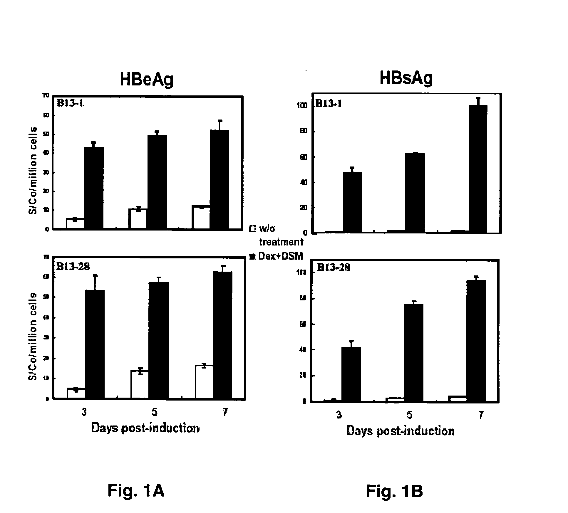
[0036] Media from B13-1 and B13-28 cells were collected on days 3, 5, and 7 after induction and were subjected to ELISA assay for both HBsAg and e antigen (Tai et al., 2002) (FIG. 1). Total cell number counts from each dish were used for normalization of the ELISA readings. As shown in FIG. 1A, e antigen secretion after induction in both B13-1 and B13-28 cells were increased by approximately 8 fold on day 3 and 4-5 fold on day 7. Surprisingly, HBsAg titer was increased by 40- to 100-fold upon induction in both B13-1 and B13-28 cells (FIG. 1B). The 2-fold increase of HBsAg from day 3 to day 7 probably reflected a doubling of cells converted to hepatocyte-like cells (FIG. 1B).
example 3
Confocal Immunofluorescence Staining of HBV Core Antigen (HBcAg) and a Liver Marker
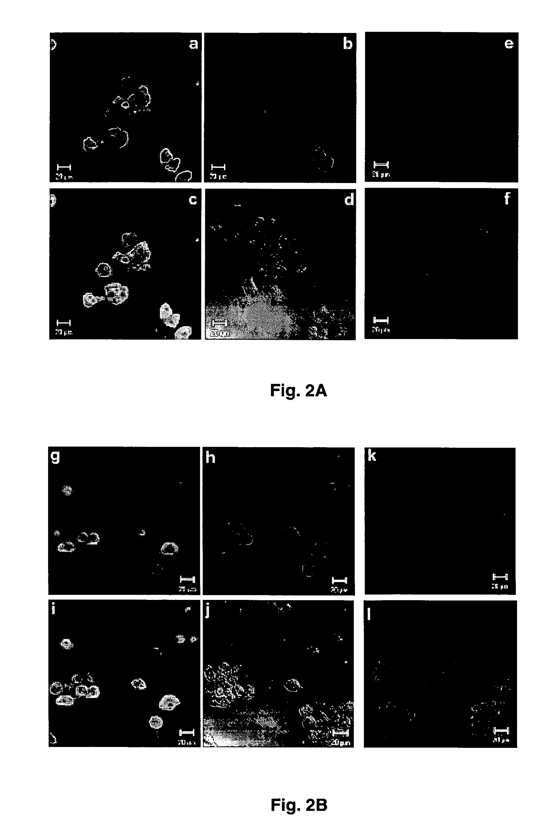
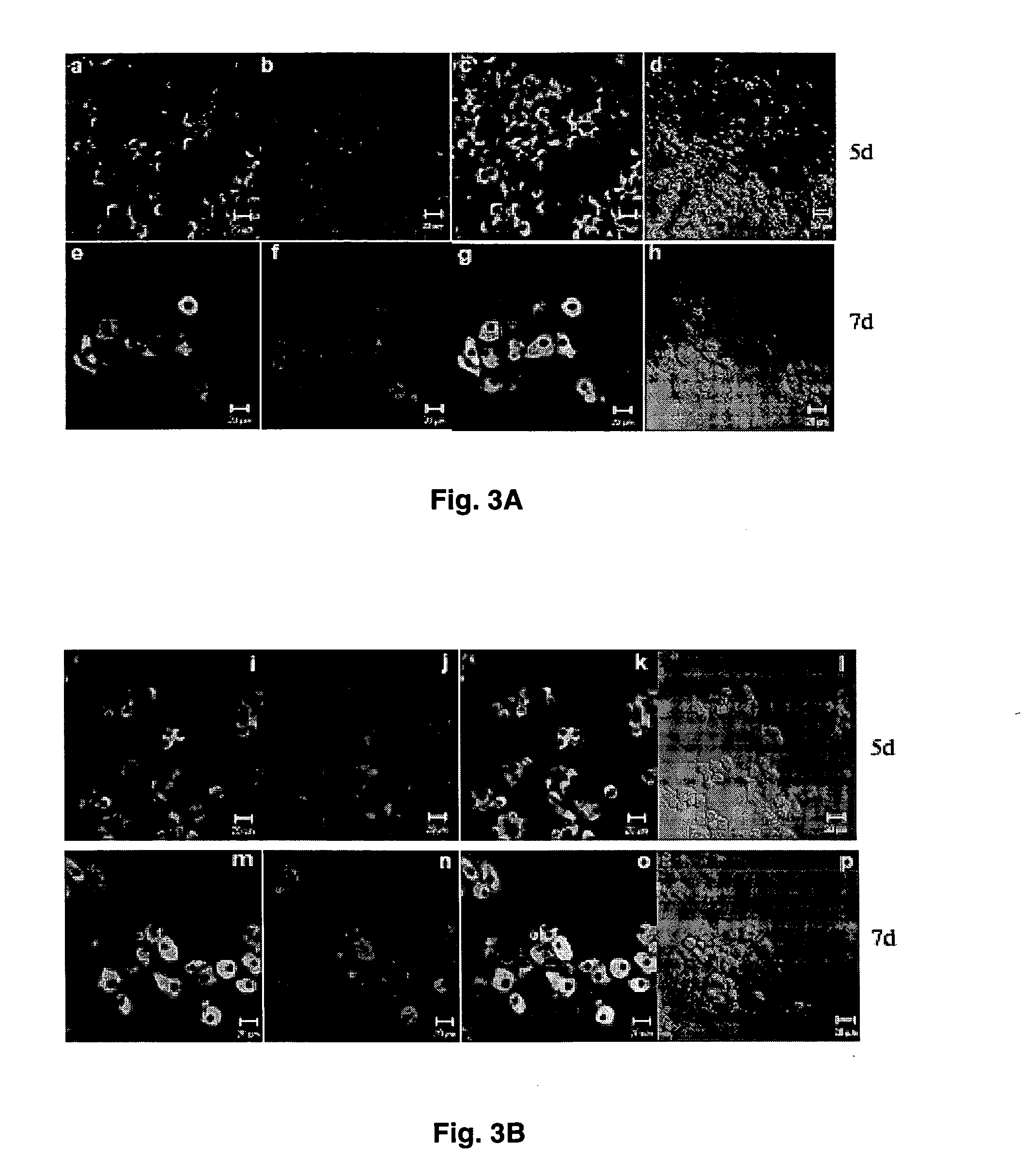
[0037] Intracellular expression of HBV core antigen (HBcAg) and liver specific glutamine synthetase were examined in B13-1 (FIG. 2A) and B13-28 cells (FIG. 2B).
[0038] For immunofluorescent staining, cells were cultured on noncoated glass coverslips, rinsed with PBS twice, fixed with 4% paraformaldehyde in PBS for 30 min at room temperature, then permeabilized with 0.1% (v / v) Triton X-100 in PBS for 30 min and incubated in 2% blocking buffer (Roche) for 1 hr. The cells were then incubated sequentially with primary and secondary antibodies (Table 1). Coverslips were incubated with DAPI (500 ng / ml in PBS) for 5 min at room temperature. After immunostaining, the coverslips were mounted on slides in gelvatol medium (20% polyvinyl alcohol in 10 mM Tris-HCl, pH 8.6). Images were collected using a Zeiss confocal microscope (LSM 510) and processed with PHOTOSHOP.
[0039] As expected, un-induced cells did not...
PUM
 Abstract
Description
Claims
Application Information
 - R&D
- Intellectual Property
- Life Sciences
- Materials
- Tech Scout
- Unparalleled Data Quality
- Higher Quality Content
- 60% Fewer Hallucinations
Browse by: Latest US Patents, China's latest patents, Technical Efficacy Thesaurus, Application Domain, Technology Topic, Popular Technical Reports.
© 2025 PatSnap. All rights reserved.Legal|Privacy policy|Modern Slavery Act Transparency Statement|Sitemap|About US| Contact US: help@patsnap.com


