

Ultrasound guided vascular access training device
- Summary
- Abstract
- Description
- Claims
- Application Information
AI Technical Summary
Benefits of technology
Problems solved by technology
Method used
Image

Examples
example 1
Construction of the Model Vessels
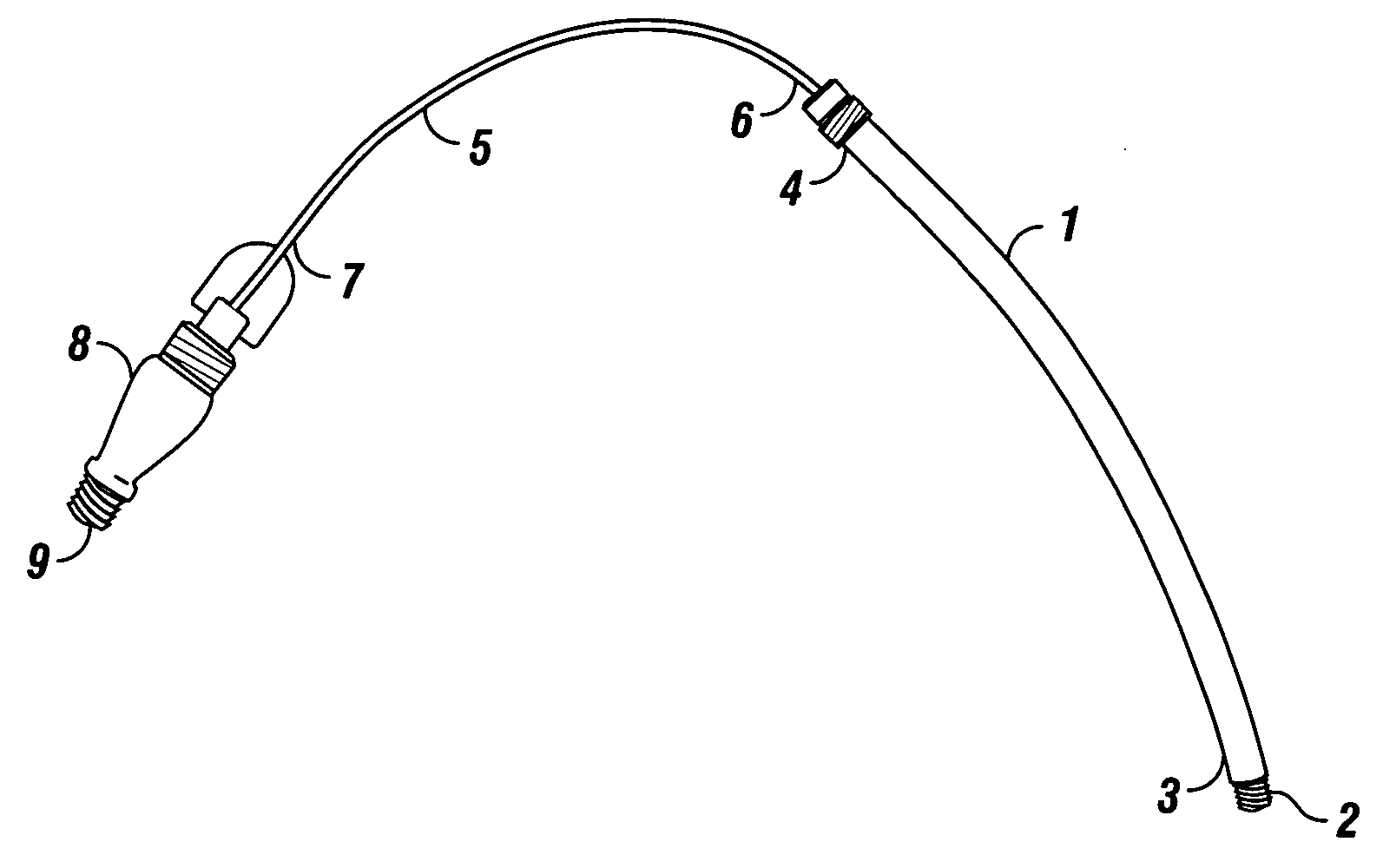
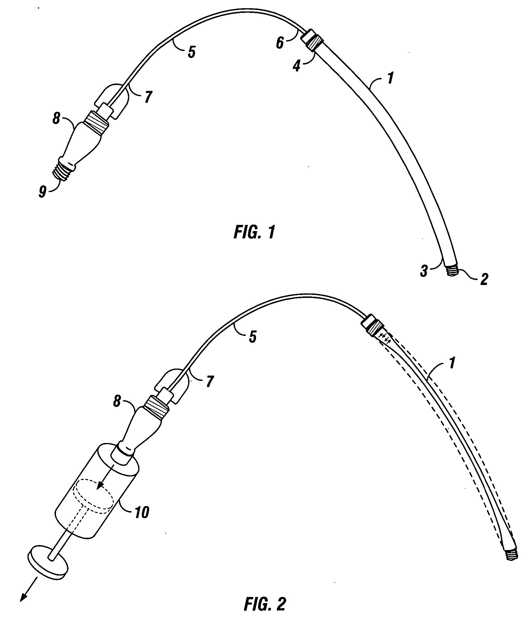
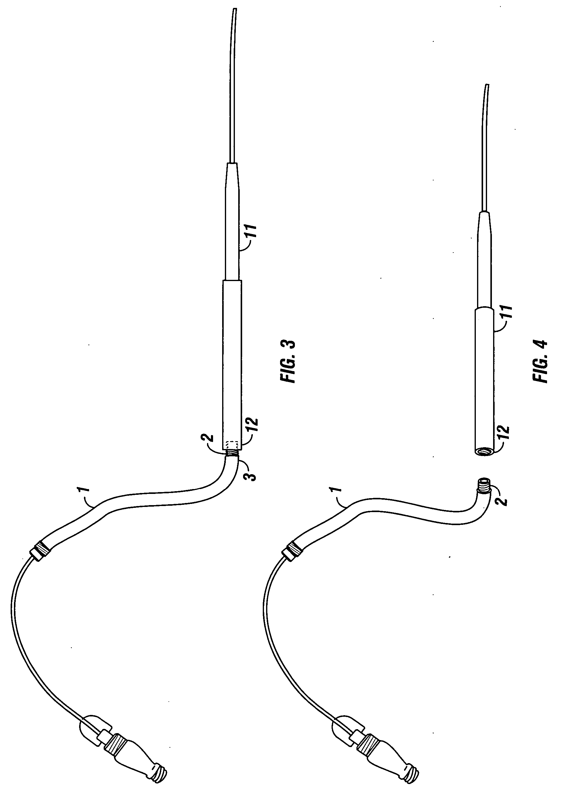
[0029] Flexible tubing, such as surgical draining tubing, selected at a thickness of 0.010 cm to 0.015 cm for a model vein, and of 0.020 cm to 0.060 cm for a model artery, is cut to a desired length, such as 9 inches for insertion into a whole chicken tissue model. The diameter of the tubing depends on the particular vascular simulation. For human neck or femoral vasculature modeling, suitable diameters range from 3 / 16 inches to ⅜ inches. One important consideration in selecting the tubing is that the material be both pliant and yet firm enough to withstand repeat puncture. Thus, natural rubber latex is a preferred material because of its elasticity and comparative ability to self-seal after puncture. Once the tubing has been cut to the desired length, one end (distal) of the tubing is sealed closed. This can be accomplished by tying the vessel end around a plug adaptor made of a rigid material, such as plastic, sized to fit into the vessel lumen. T...
example 2
Insertion of the Model Vasculature into a Tissue Model
[0030] A tissue model, preferably containing anatomical structures, such as bones, is chosen for the insertion of the model vasculature. As a convenient example, a whole chicken may be used. However, for veterinary applications, for example, a larger tissue model, such as a turkey or a pig, may be preferable. The inclusion of anatomical structures provides the trainee with a realistic training model for ultrasound-guided vascular access procedures. The fully assembled model vasculature comprising at least one model artery and one model vein is air-evacuated in preparation for insertion into the tissue model. Air evacuation serves to reduce the diameter of the vessel and to minimize the hole created in the tissue model. A standard syringe is threaded onto the two-way valve located at the end of the connecting tubing that is distal to the model vessel and air is withdrawn from the model vessel lumen. Once the air has been removed ...
example 3
[0033] Ultrasound-Guided Vascular Access Training
[0034] Once the training vasculature is in place and has been loaded with fluid, trainees can begin to practice ultrasound-guided vascular access techniques. For this purpose, a hand-held ultrasound device is applied to the surface of the vascularized tissue model and the trainee locates the model vessels on the sonographic image displayed on a screen. After locating the vessels, the trainee can distinguish a model artery from a model vein by pressing down onto the tissue with the hand-held ultrasound device. The model artery will be relatively resistant to the pressure and retain its circular shape, while the model vein will deform more readily upon application of pressure. While still holding the ultrasound device and monitoring the sonographic image, the trainee can then insert a needle into the tissue model. Because the needle and its tip are visible on ultrasound, the trainee can visually follow its path as he / she guides it towa...
PUM
 Abstract
Description
Claims
Application Information