Low coherence interferometry for detecting and characterizing plaques

a low coherence, plaque technology, applied in the field of low coherence interferometry for detecting plaques, can solve the problems of large number of victims of chd who are apparently healthy and die suddenly, insufficient screening and diagnostic methods, and vulnerable plaques buried inside the arterial wall, etc., to facilitate the interference of broadband light reflected

Inactive Publication Date: 2005-11-17
VZN CAPITAL
View PDF99 Cites 151 Cited by
  • Summary
  • Abstract
  • Description
  • Claims
  • Application Information

AI Technical Summary

Problems solved by technology

Despite major advances in the treatment of coronary heart disease patients, a large number of victims of CHD who are apparently healthy die suddenly without prior symptoms.
Available screening and diagnostic methods are insufficient to identify the victims before the catastrophic event occurs.
In fact, vulnerable plaque may be buried inside the arterial wall.
When this thin covering over the plaque cracks and bleeds, it spills the contents of the vulnerable plaque into the bloodstream, creating clots large enough to block the artery.
This narrowing or clogging interferes with the normal flow of blood, sometimes causing pain but often exhibiting no symptoms at all.
In some cases, PVD may be caused by blood clots that lodge in the arteries and restrict blood flow.
In extreme cases, untreated PVD can lead to gangrene, a serious condition that may require amputation of a leg, foot or toes.
While the fibrous tissue within the cap provides structural integrity to the plaque, the interior of the atheroma is soft, weak and highly thrombogenic.
However, angiography is only sensitive enough to detect hemodynamically significant lesions (>70% stenosis), which account for approximately 33% of acute coronary syndrome cases.
Because angioscopic visualization is limited to the surface of the plaque, it is generally insufficient for use in detecting actively forming atheromatous plaques and / or determining vulnerable plaques.
IVUS can detect thin caps and distinguish regions of intermediate density (e.g., intima that is rich in smooth muscle cells and fibrous tissue) from echolucent regions, but current technology does not determine which echolucent regions are composed of cholesterol pools rather than thrombosis, hemorrhage, or some combination thereof.
However, thermography may not provide information about eroded but non-inflamed lesions, vulnerable or otherwise, having a propensity to rupture.
However, it is unlikely that actively forming and / or vulnerable plaques will be reliably differentiated from stable plaques based solely on this ratio.
Unfortunately, OCT is an imaging technique and, as a result, is computationally intensive and very time consuming.
Unfortunately, current LCI techniques, such as OCT, rely on amplitude measurements of the interferences signal and may lack the high resolution required for accurate detection and characterization of vulnerable plaques.
In spite of these endeavors, attempts to make available an effective sensor for practical operation to detect vulnerable plaque have thus far, proved inadequate.

Method used


Image

  • Low coherence interferometry for detecting and characterizing plaques
  • Low coherence interferometry for detecting and characterizing plaques
  • Low coherence interferometry for detecting and characterizing plaques

Examples


Embodiment Construction

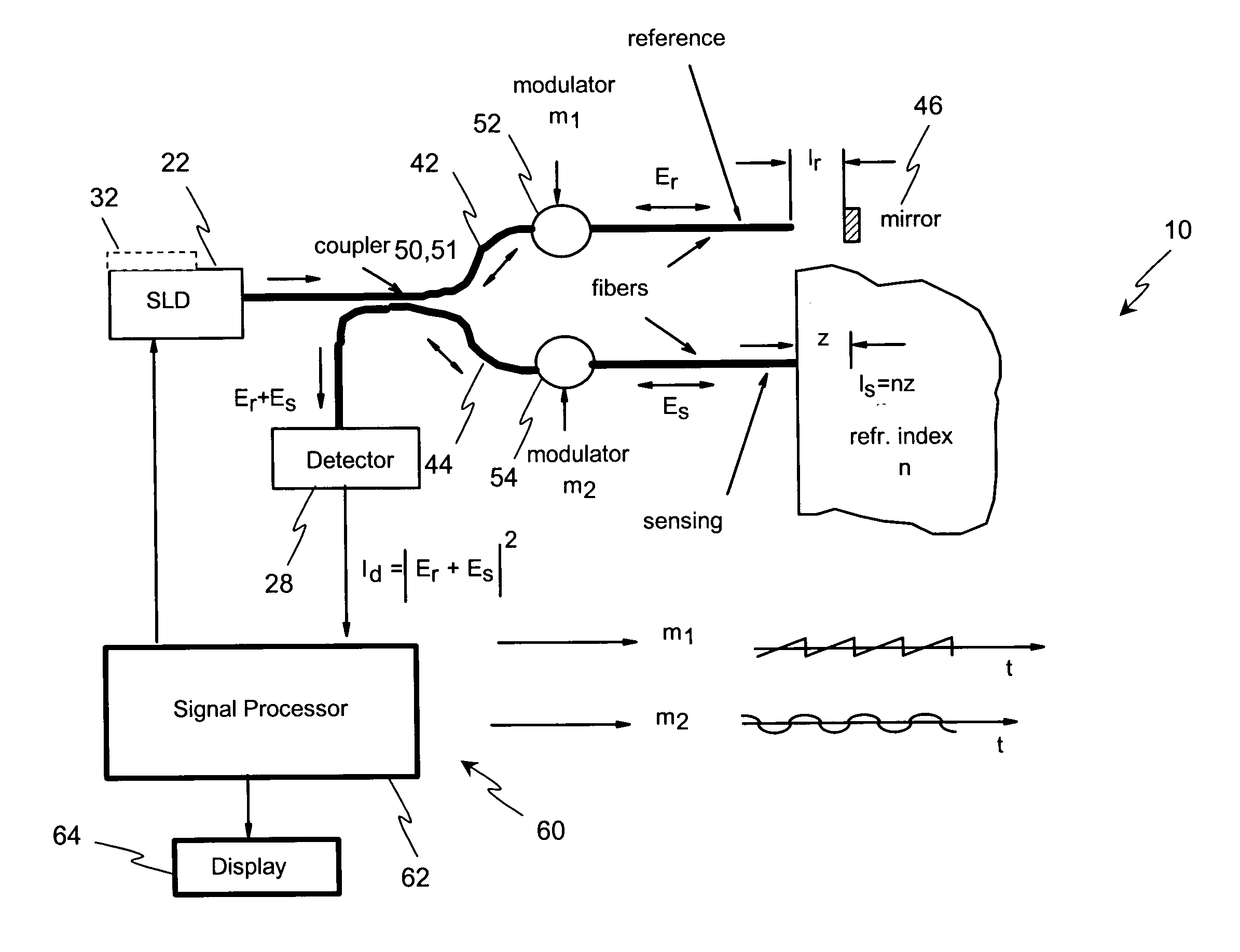
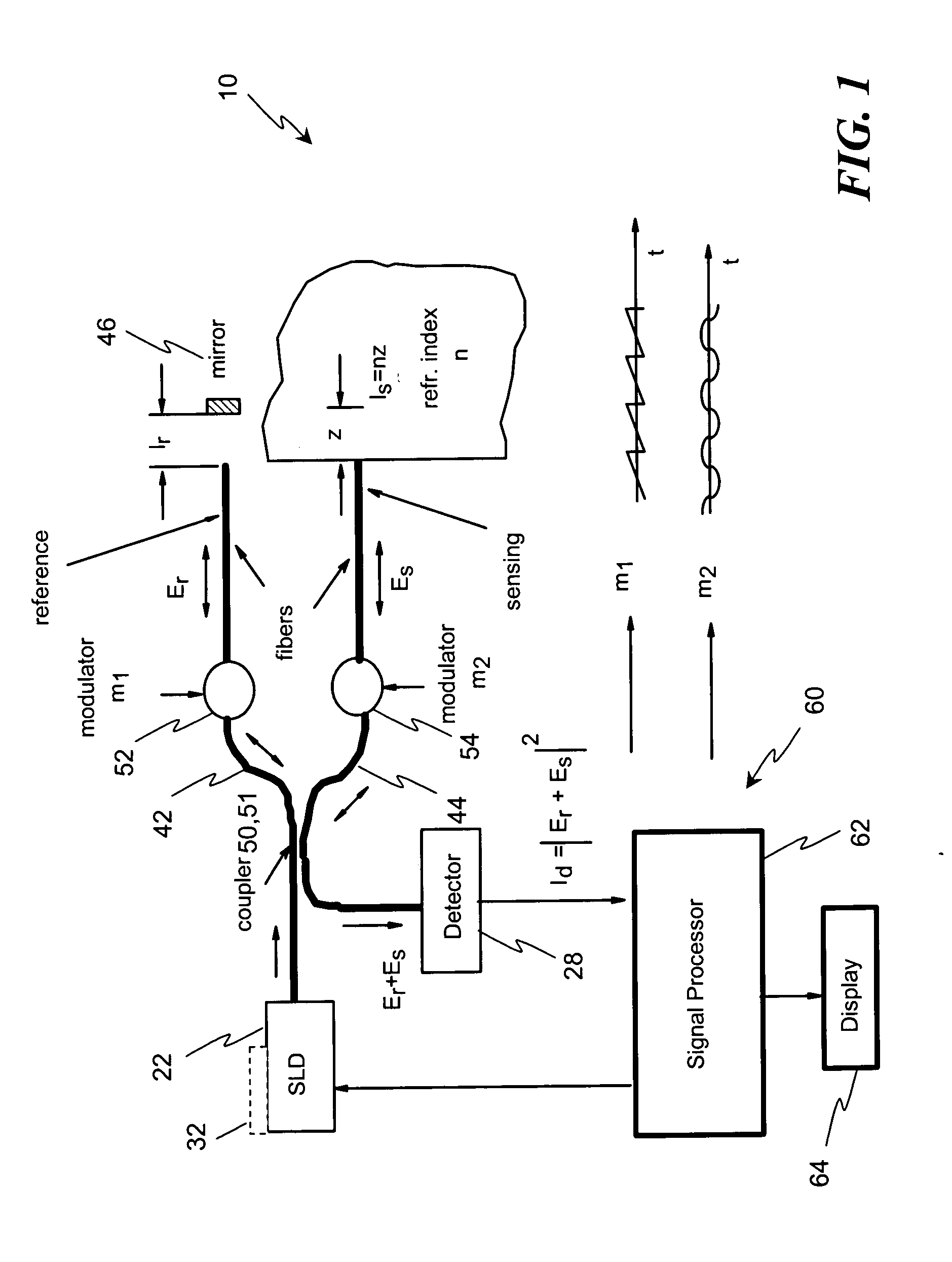
[0064] Disclosed herein, in several exemplary embodiments are high-sensitivity low coherence interferometric (LCI) systems (instruments) for optical metrology, for use in a variety of sensing and monitoring applications, including, but not limited to, trace chemical sensing, optical properties and changes thereof, medical sensing such as detecting and characterizing vulnerable plaques and others. In an exemplary embodiment, the instrument is miniaturized, using integrated optics components such as waveguides, splitters and modulators on a single substrate such as, but not limited to, a LiNbO3 (Lithium Niobate) chip. The exemplary embodiments may also involve the use of a “circulator” type of optical component, including of a polarizing beam splitter and quarterwave plate, which can be combined with the light source and detector into a miniature module that prevents optical feedback into the light source while doubling the detected light. Alternatively, instead of the polarizing beam...


PUM

PropertyMeasurementUnit
critical thicknessaaaaaaaaaa
light wavelengthsaaaaaaaaaa
wavelengthaaaaaaaaaa

Abstract

A method and system for determining a characteristic of a biological sample including directing light at the biological sample and receiving that light; directing the light at a reference reflecting device and receiving that light; adjusting an effective light path length to facilitate an interference of the light reflected from the biological sample corresponding to a first depth and the light reflected from the reference reflecting device; and detecting the broadband light resulting from the interference, to provide an interference signal. The method also includes: determining a first phase associated with the interference signal corresponding to the first depth; varying the effective light path length to define a second depth; determining a second phase associated with the interference signal corresponding to the second depth; and determining the characteristic of the biological sample from the phases.

Description

CROSS-REFERENCE TO RELATED APPLICATIONS [0001] This is a continuation-in-part application of U.S. Ser. No. 10 / 845,849, filed May 14, 2004 the contents of which are incorporated by reference herein in their entirety.BACKGROUND [0002] The invention concerns a low coherence interferometric (LCI) method for detecting plaques. For example, a method and apparatus for the analysis and detection of atheromas and / or atherosclerotic plaques in arterial walls. More particularly, in one embodiment, a methodology and system for detecting and evaluating vulnerable atherosclerotic plaques in a blood vessel. Further, measurement of the interface between the plaque and lipid pool between the plaque and the artery and measurement the thickness of the plaque with a high accuracy. An application area of interest is that of the diagnosis and management of cardiovascular diseases (CVD). [0003] Coronary Heart Disease (CHD) accounts for approximately fifty percent of the death toll attributed to CVD. Despi...

Claims


Application Information

Patent Timeline
Patent Type & Authority Applications(United States)
IPC IPC(8): A61B5/00G01B9/02G01N21/45G01N21/47G01N21/49
CPCA61B5/0066A61B5/0086A61B5/02007A61B5/14532A61B5/14546G01N21/49A61B2562/0242G01N21/45G01N21/474G01N21/4785A61B5/1455
Inventor ALPHONSE, GERARD A.
Owner VZN CAPITAL
Why Eureka
Try Eureka