Function of autophagy genes in cell death

Inactive Publication Date: 2005-12-15
NAT INST OF HEALTH REPRESENTED BY THE SEC OF THE DEPT OF HEALTH & HUMAN SERVICES NAT INST OF HEALTH +1
View PDF1 Cites 22 Cited by
  • Summary
  • Abstract
  • Description
  • Claims
  • Application Information

AI Technical Summary

Benefits of technology

[0014] In still another aspect, the present invention relates to a method for inhibiting cell death in a mammalian cell, the method comprising reducing expression of receptor interacting protein (RIP) and inhibiting caspase 8 in the mammalian cell.
[0015] Another aspect of the present invention relates to a method for inducing and increasing autophagic cell death in mammalian cells, the method comprising reducing expression of caspase 8 and increasing expression of ATG7 (HsGSA7/mAPG7) or Beclin 1.
[0016] Yet, another aspect of the present invention relates to a method for increasing cell death by the autophagy pathway, the method comprising increasing express

Problems solved by technology

Necrosis can result when cell metabolism and integrity are compromised by a nonphysiological insult.
The pro

Method used


Image

  • Function of autophagy genes in cell death
  • Function of autophagy genes in cell death
  • Function of autophagy genes in cell death

Examples


example 1

Autophagy and ATG Genes are Required for zVAD-Induced Cell Death.

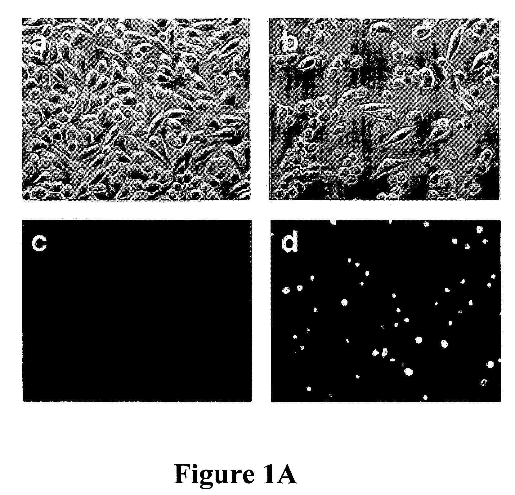
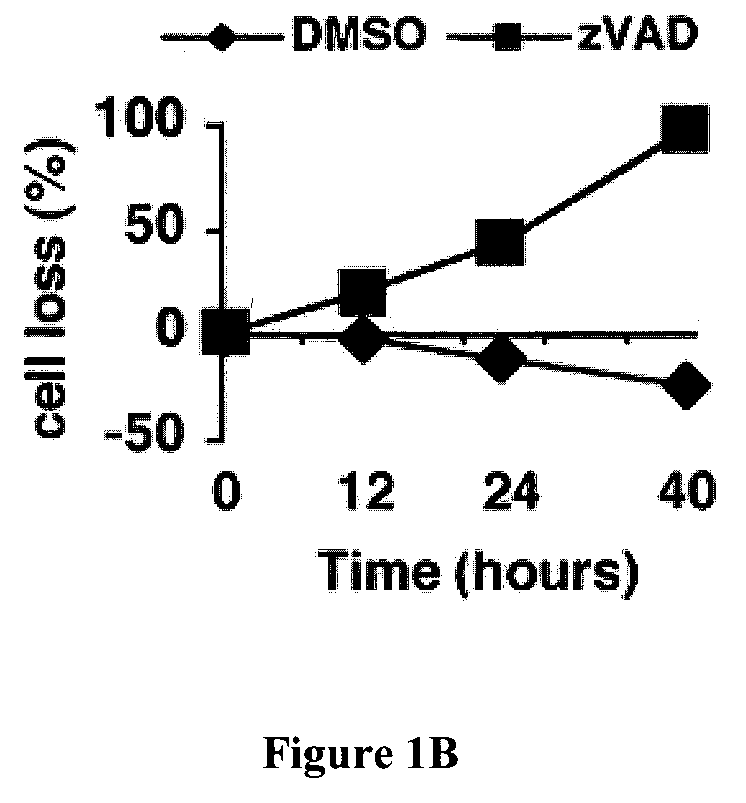
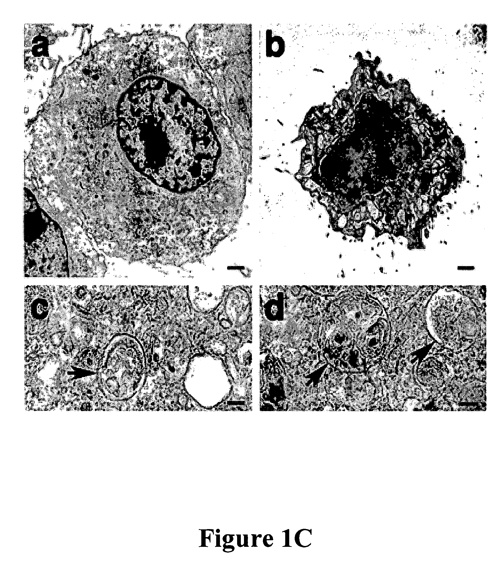
[0098] In mouse L929 fibroblastic cells, tumor necrosis factor (TNF), oxidants, ceramide, and radiation can induce caspase-independent death (11). However, benzyloxycarbonyl-Valyl-Alanyl-Aspartic-acid (O-methyl)-fluoromethylketone (zVAD), a caspase inhibitor with broad specificity, also directly induced the death of L929 cells. L929 cells were treated with 1 ul of dimethyl sulfoxide (DMSO) FIG. 1A (a and c) or 20 uM zVAD (b and d) for 24 hours and examined by phase contrast microscopy (a and b); or 4′,6′-diamidino-2-phenylindole-staining and fluorescent microscopy (c and d). Magnification: 200×. Death began at 12 hours after zVAD treatment and was complete after 40 hrs as shown in FIG. 1B. Transmission electron microscopy (TEM) revealed intact mitochondria and endoplasmic reticulum, condensed osmophilic cytoplasm, and numerous large cytoplasmic inclusions that were membrane-bound vacuoles characteristic of autophagy ...

example 2

Requirement of RIP and JHK Signaling Pathways for Autophagic Death

[0106] Death receptors can elicit nonapoptotic death through the “receptor-interacting protein” (RIP), a death-domain-containing, serine-threonine kinase (6, 7). RIP expression was reduced by RNAi and decreased autophagy and cell death was observed. For example, cells were treated with zVAD after transfection with RIP RNAi or nonspecific (NS) oligonucleotides. Reduction in cell number (solid bars) and the fractions of cells with autophagic features based on TEM (open bars) are shown in FIG. 2A. FIG. 2B is a western blot for phospho-JNK (left lane) or total JNK protein (right lane) after zVAD treatment. zVAD activated c-Jun N-terminal kinase (JNK) that is also activated by RIP in response to cytokines.

[0107] L929 cells were pretreated with 1 ug / ml JNK inhibitor II, 1 ug / ml p38 inhibitor SB 203580, or 1 ug / ml Erk inhibitor PD 98059 for 1 hour, and were then treated with 20 uM zVAD for 40 hours, with zVAD dissolved in...

example 3

Inhibition of the Autophagic Death Pathway by Caspase-8

[0109] Finally, the mechanism as to how zVAD induced autophagic cell death was investigated. RNAi was used to progressively reduce caspase-8 expression over time. FIG. 3A shows the time course of viability of L929 cells transfected with either nonspecific (NS) (open bars) or caspase 8-specific (solid bars) RNAi at 24, 96, and 110 hours after transfection. It was found that cell death was correspondingly increased by inhibiting the expression of caspase 8 and thereby inducing the autophagic pathway. Inset panels show the abundance of caspase-8 protein by Western blot.

[0110] Representative TEM pictures and quantification of the cells treated with either nonspecific or caspase-8 specific RNAi showed features of autophagy. Cells were harvested at 96 hours after RNAi transfection (a) NS control cell, (b to d) Caspase-8 RNAi at different magnifications. The arrows in c and d show double-membrane autophagic vacuoles. FIG. 3B(e) show...


PUM

PropertyMeasurementUnit
Fractionaaaaaaaaaa
Cell deathaaaaaaaaaa
Morphologyaaaaaaaaaa

Abstract

The present invention relates to a new molecular pathway in which activation of the receptor-interacting protein (RIP, a serine-threonine kinase) and Jun N-terminal kinase induce cell death with the morphology of autophagy. Further, autophagic death is induced by caspase 8 inhibition and expression of the mammalian genes ATG7 and beclin.

Description

CROSS REFERENCE TO RELATED APPLICATIONS [0001] This application claims the benefit from U.S. Provisional Application Ser. No. 60 / 566,857, filed Apr. 30, 2004, the content of which is hereby incorporated by reference herein for all purposes.BACKGROUND OF THE INVENTION [0002] 1. Field of the Invention [0003] The present invention relates generally to cell death, and more particularly, to methods and compositions for regulating cell death through activating the autophagic pathway. [0004] 2. Background of the Related Art [0005] Apoptosis is a well-studied pathway of programmed cell death conserved from C. elegans to humans (1). Caspases, a family of cystinyl, aspartate-requiring proteases produce the morphological changes associated with apoptotic death (2, 3). Non-apoptotic forms of cell elimination include those with features of necrosis and autophagy (4-7). Autophagy is a process that liberates free amino acids and nucleotides and enables cells to survive under nutrient deprivation c...

Claims


Application Information

Patent Timeline
IPC IPC(8): A61K38/05A61K38/06A61K39/395A61K48/00C12N15/113
CPCA61K38/06C12N15/1137C07K14/8139C12Y304/22056C12Y304/22061C12N2310/14
Inventor BAEHRECKE, ERICALVA, AJJAILENARDO, MICHAELLI, YU
Owner NAT INST OF HEALTH REPRESENTED BY THE SEC OF THE DEPT OF HEALTH & HUMAN SERVICES NAT INST OF HEALTH
Why Eureka
Try Eureka