Inhibition of histone deacetylase as a treatment for cardiac hypertrophy

a technology of histone deacetylase and cardiac hypertrophy, which is applied in the field of development biology and molecular biology, can solve the problems of cardiac hypertrophy, which is still not fully understood, and achieve the effects of improving one or more symptoms of cardiac failure, increasing exercise capacity, and increasing blood ejection volum

Inactive Publication Date: 2006-02-02
LONG CARLIN +3
View PDF7 Cites 2 Cited by
  • Summary
  • Abstract
  • Description
  • Claims
  • Application Information

AI Technical Summary

Benefits of technology

[0010] Thus, in accordance with the present invention, there is provided a method of treating pathologic cardiac hypertrophy and heart failure comprising (a) identifying a patient having cardiac hypertrophy; and (b) administering to the patient a histone deacetylase inhibitor. Administering may comprise intravenous, oral, transdermal, sustained release, suppository, or sublingual administration. The method may further comprise administering a second therapeutic regimen, such as a beta blocker, an iontrope, diuretic, ACE-I, AII antagonist or Ca-blocker. The second therapeutic regimen may be administered at the same time as the histone deacetylase inhibitor, or either before or after the histone deacetylase inhibitor. The treatment may improve one or more symptoms of cardiac failure such as providing increased exercise capacity, increased blood ejection volume, left ventricular end diastolic pressure, pulmonary capillary wedge pressure, cardiac output, cardiac index, pulmonary artery pressures, left ventricular end systolic and diastolic dimensions, left and right ventricular wall stress, wall tension and wall thickness, quality of life, disease-related morbidity and mortality.

Problems solved by technology

All of these signals activate MEF2 and result in cardiac hypertrophy.
However, it is still not completely understood how the various signal systems exert their effects on MEF2 and modulate its hypertrophic signaling.

Method used


Image

  • Inhibition of histone deacetylase as a treatment for cardiac hypertrophy
  • Inhibition of histone deacetylase as a treatment for cardiac hypertrophy
  • Inhibition of histone deacetylase as a treatment for cardiac hypertrophy

Examples


example 1

Materials and Methods

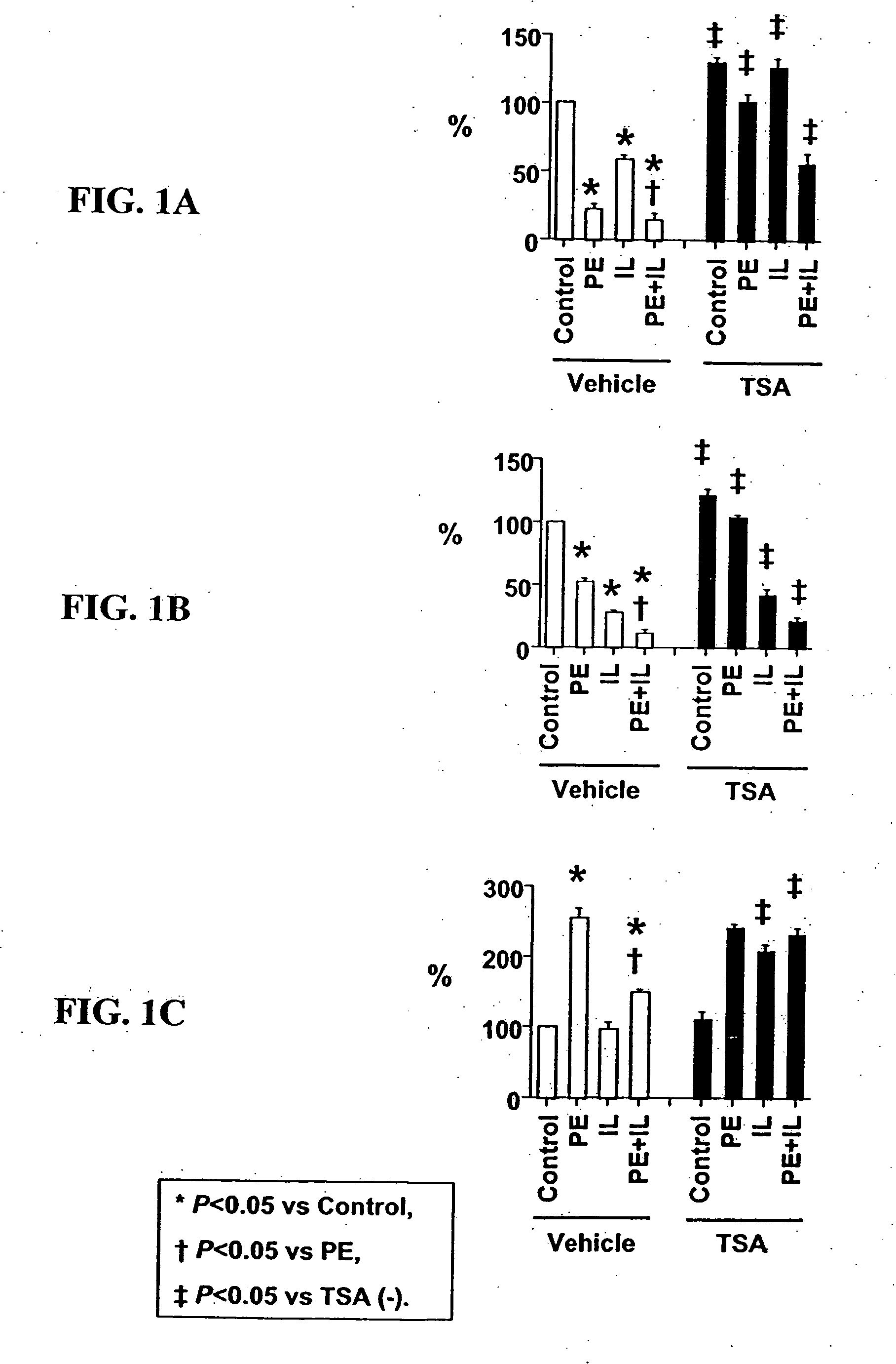
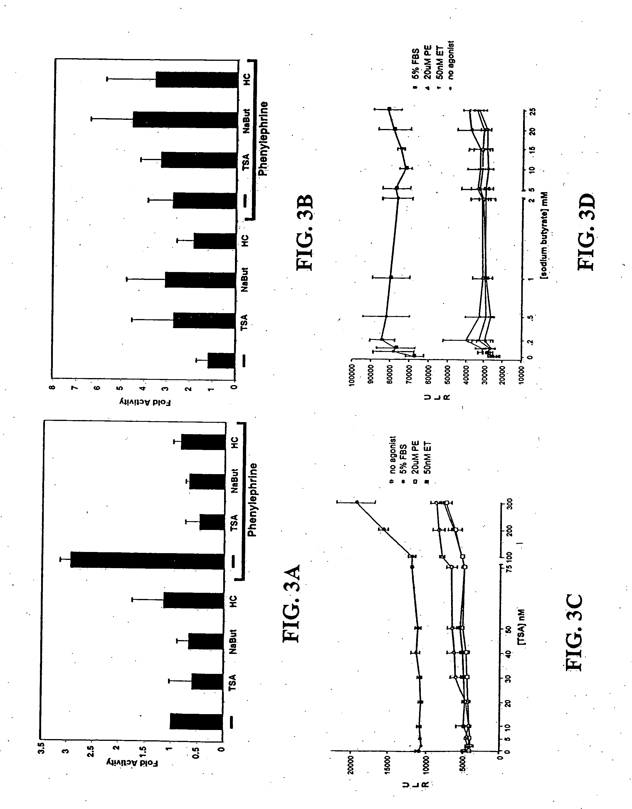
[0099] Cell culture. Ventricular myocytes from one-day-old rats were plated at low density in MEM with 5% calf serum, and studied in serum-free MEM. Cultures were treated with PE (#P6126, Sigma-Aldrich Corp., St. Louis, Mo.), IL-1 (#501-RL, R&D Systems, Minneapolis, Minn.), TSA (#GR-309, BIOMOL Research Laboratories, Inc., Plymouth Meeting, Pa.) or their vehicles (ascorbic acid for PE; bovine serum albumin for IL-1; dimethyl sulfoxide for TSA).

[0100] Detection of acetylated lysine residues. Total cell extract was subjected to Western blot analysis using antibodies from Cell Signaling Technology (Beverly, MA) (acetylated lysine antibody: #9441, acetylated histone H3 antibody at Lysine 9: #9671, acetylated histone H3 antibody at Lysine 23: #9674, histone H3 antibody: #9712). Nuclear localization of acetylated histone H3 was examined by immunostaining using the same antibodies.

[0101] Quantification of myocyte hypertrophy and myocyte-specific mRNA expression. Gr...

example 2

Materials and Methods

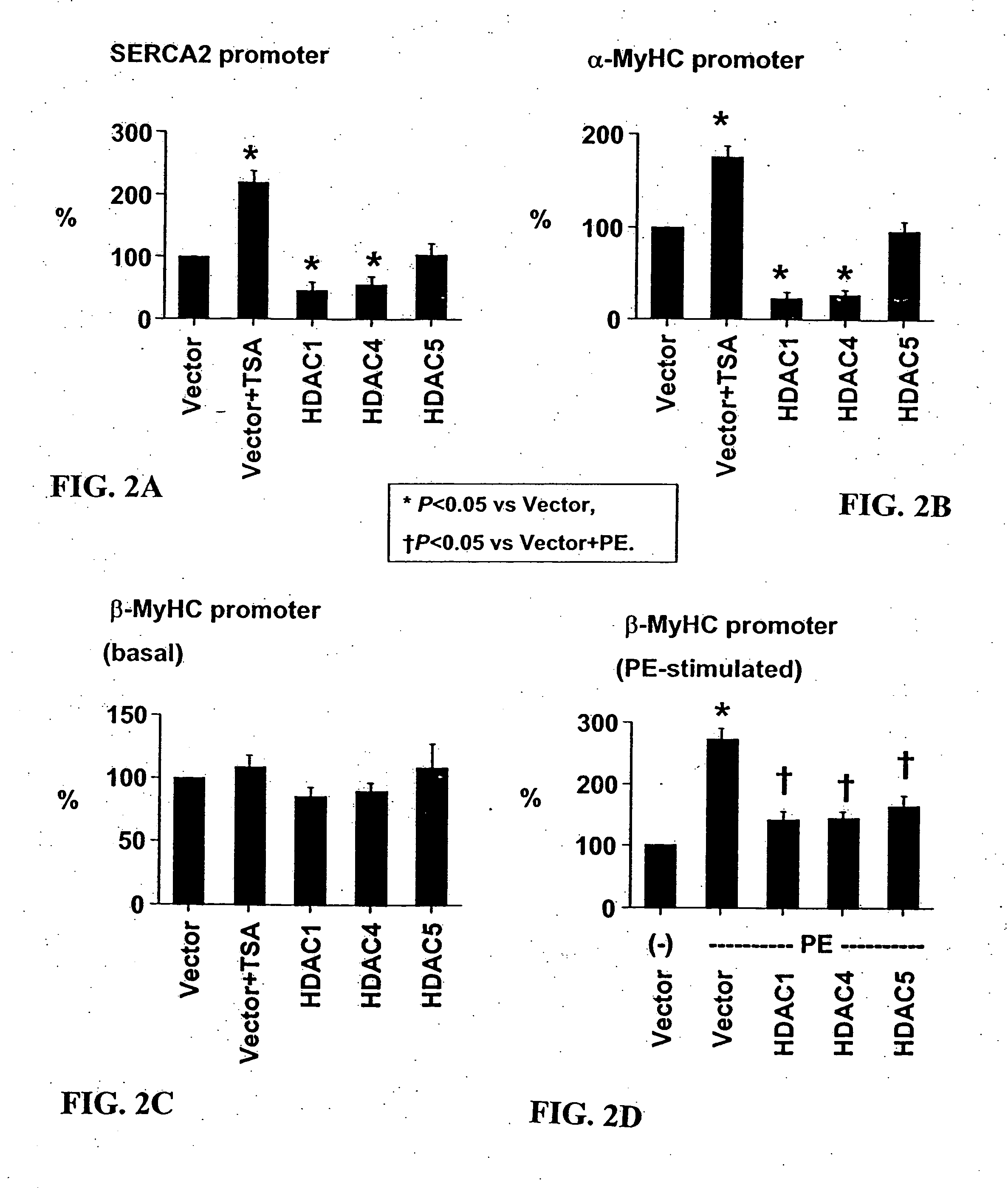
[0106] Cardiomyocyte isolation. Hearts were harvested from 15-day timed-pregnant female Sprague-Dawley rats (Harlan, Houston, Tex.). After mincing in phosphate-buffered saline, cardiomyocytes were isolated from successive digestion fractions of 0.1% (w / v) Pancreatin (Sigma, St. Louis, Mo.) solution. Fractions were collected; resuspended in plating medium, pooled; and then plated for 2 hours to separate fibroblasts from cardiomyocyte population. Suspended cells were recollected and plated in 6-well dishes at 1×10⁶ cells / well for transfection and immunofluorescence experiments, and 2×10⁶ cells in 10 cm dishes for RNA analysis.

[0107] Transcription assay. Twenty-four hours after plating, cells were transfected with total of 1 μg / ml for 5 hrs using Lipofectamine Plus reagent (Invitrogen, Carlsbad, Calif.). Transfected cells were incubated 24 hrs then treated for an additional 24 hrs. Cells were lysed and their lysates assayed with the Luciferase Assay System (Prom...


PUM

PropertyMeasurementUnit
cell sizeaaaaaaaaaa
massaaaaaaaaaa
timeaaaaaaaaaa

Abstract

The present invention provides for methods of treating and preventing cardiac hypertrophy. Class II HDACs, which are known to participate in regulation of chromatin structure and gene expression, have been shown to have beneficial effects on cardiac hypertrophy. Surprisingly, the present invention demonstrates that HDAC inhibitors inhibit cardiac hypertrophy by inhibiting fetal cardiac gene expression and interfering with sarcomeric organization.

Description

[0001] The present invention claims benefit of priority to U.S. Provisional Ser. Nos. 60 / 325,311, filed Sep. 27, 2001, and 60 / 334,041, filed Oct. 31, 2001, the entire contents of which are hereby incorporated by reference without reservation.[0002] The government owns rights in the present invention pursuant to grant number NIH RO1 HL61544 from the National Institutes of Health.BACKGROUND OF THE INVENTION [0003] 1. Field of the Invention [0004] The present invention relates generally to the fields of developmental biology and molecular biology. More particularly, it concerns gene regulation and cellular physiology in cardiomyocytes. Specifically, the invention relates to the use of HDAC inhibitors to treat cardiac hypertrophy and heart failure. [0005] 2. Description of Related Art [0006] Cardiac hypertrophy in response to an increased workload imposed on the heart is a fundamental adaptive mechanism. It is a specialized process reflecting a quantitative increase in cell size and mas...

Claims


Application Information

Patent Timeline
Patent Type & Authority Applications(United States)
IPC IPC(8): A61K38/12A61K31/19A61K31/16A61K31/165A61K31/167A61K31/18A61K31/336A61K31/40A61K31/4402A61K31/473A61K38/00A61K45/00A61K45/06A61P9/00A61P9/04G01N33/15G01N33/50
CPCA61K31/165A61K31/19A61K31/401A61K38/12A61K45/06A61K2300/00A61P43/00A61P9/00A61P9/04A61P9/10A61P9/12
Inventor LONG, CARLINOLSON, ERICBRISTOW, MICHAELMCKINSEY, TIMOTHY
Owner LONG CARLIN
Why Eureka
Try Eureka