Therapeutic uses of tim-3 modulators

a technology of tim3 and tim-3, which is applied in the field of therapeutic use of tim3 modulators, can solve the problems of no longer being remyelinated, progressive neurodegeneration, no known cure for ms, etc., and achieve the effects of increasing tim-3 activity, enhancing tnf-a secretion, and increasing tim-3 activity

Inactive Publication Date: 2010-03-11
THE BRIGHAM & WOMEN S HOSPITAL INC
View PDF1 Cites 73 Cited by
  • Summary
  • Abstract
  • Description
  • Claims
  • Application Information

AI Technical Summary

Benefits of technology

[0039]Another aspect provided includes a vaccine composition comprising an agent that increases TIM-3 activity and a TLR ligand. In one embodiment, the agent increases TIM-3 activity in an antigen presenting cell (APC). This aspect is based, in part, on the observation that TIM-3 activation enhances the secretion of TNF-a by m

Problems solved by technology

This continuous demyelination leads to more dystrophic neurons that over time can no longer be remyelinated, leading to progressive neurodegeneration.
There is presently no known cure for MS.
Glioblastomas cause death due to rapid, aggressive, and infiltrative growth in the brain.
In addition, the immune response to the neoplastic cells is mainly ineffective in completely eradicating residual neoplastic cells foll

Method used


Image

  • Therapeutic uses of tim-3 modulators
  • Therapeutic uses of tim-3 modulators
  • Therapeutic uses of tim-3 modulators

Examples


examples

Methods

Flow Cytometry

[0302]Single cell suspension from collagenase treated spleens were stained with the following antibodies: anti-CD11b, anti-CD11c, anti-TIM-3 (EBioscience) and Rat IgG1 isotype control (BD Biosciences). All data were collected on a FACSCalibur (BD Biosciences) and analyzed with FlowJo software (Tree Star).

Real-Time Quantitative PCR

[0303]Total RNA was prepared from sorted cell populations using Trizol (Invitrogen) followed by RNA clean-up and DNaseI digestion (Qiagen). RNA was then reverse transcribed to cDNA and used as template in quantitative RT-PCR using the Taqman system (Applied Biosystems). Primers and probes for detection of full-length mouse TIM-3 were designed in the mucin domain. Sequences were as follows: Probe, 5′-AGA CAC TGG TGA CCC TCC ATA ATA ACA ATG GAA-3′ (SEQ ID NO:7); Forward primer, 5-CGG AGA GAA ATG GTT CAG AGA CA-3′ (SEQ ID NO:8); Reverse primer, 5-TTC ATC AGC CCA TGT GGA AAT-3′ (SEQ ID NO:9). GAPDH primers and probes were purchased from App...

example 1

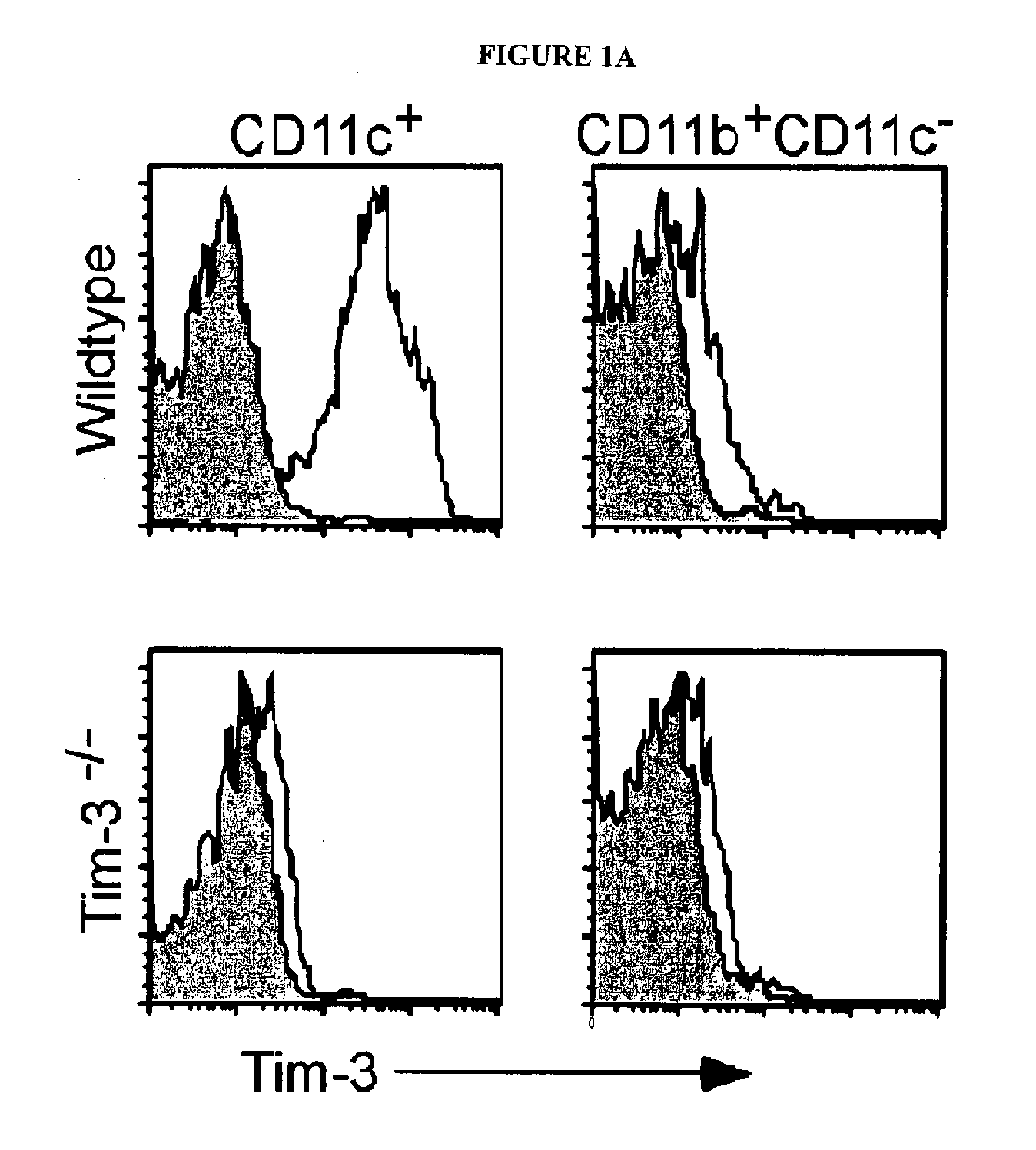
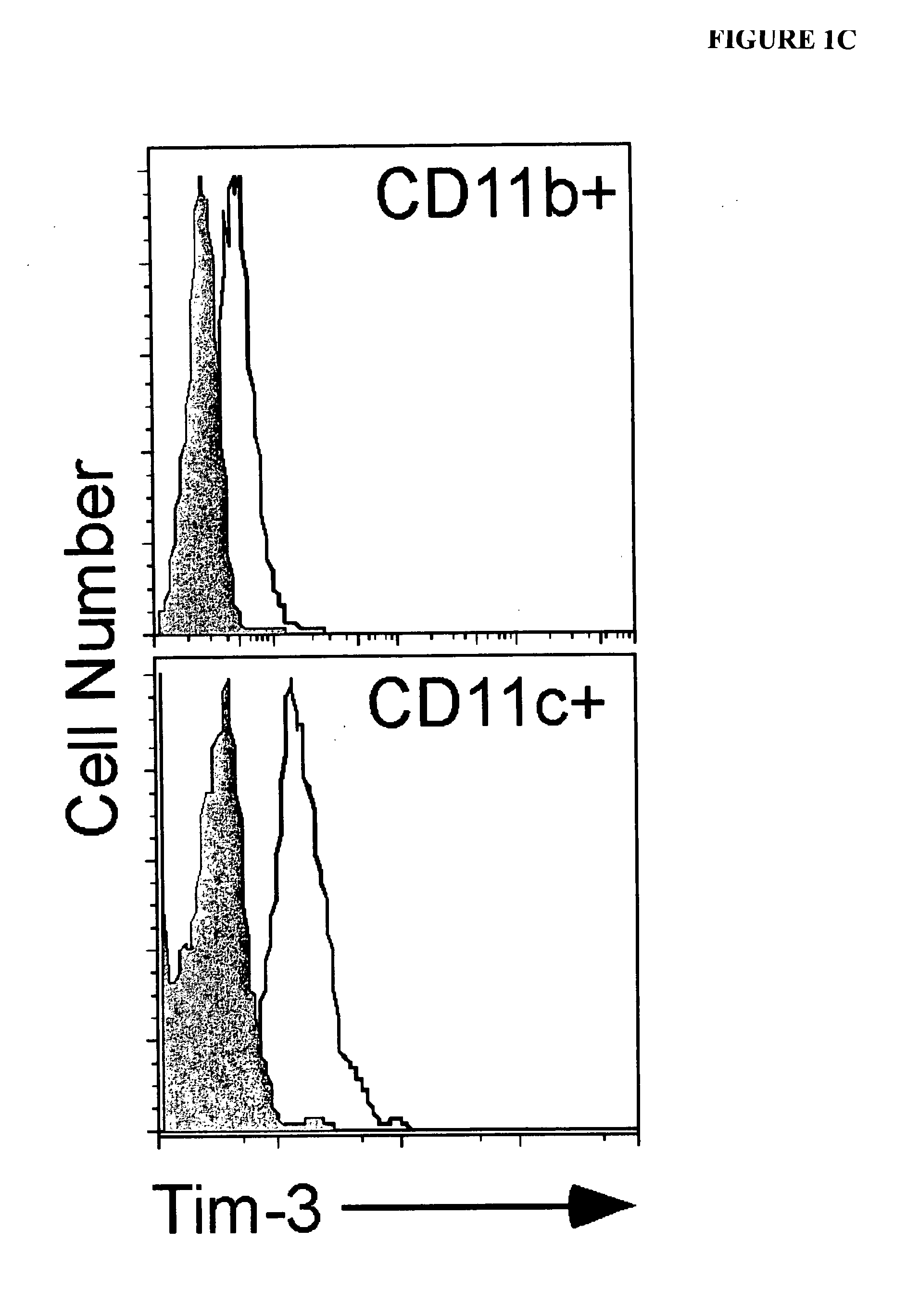
[0312]The expression of TIM-3 mRNA and protein in macrophages and myeloid dendritic cells (DCs) was examined. Murine macrophages (CD11b+CD11c−) did not express TIM-3, while nearly all CD11c+DCs expressed high levels of TIM-3 directly ex vivo (FIG. 1A). The staining of TIM-3 on DCs was specific, as no staining was observed on CD11c+ cells from TIM-3− / − mice. The expression of TIM-3 on lymphoid (CD8+), myeloid (CD11b+), and plasmacytoid (B220+) DCs was also examined. TIM-3 was equally expressed on all subsets. TIM-3 was expressed at similar levels on immature versus mature bone marrow derived DCs.

example 2

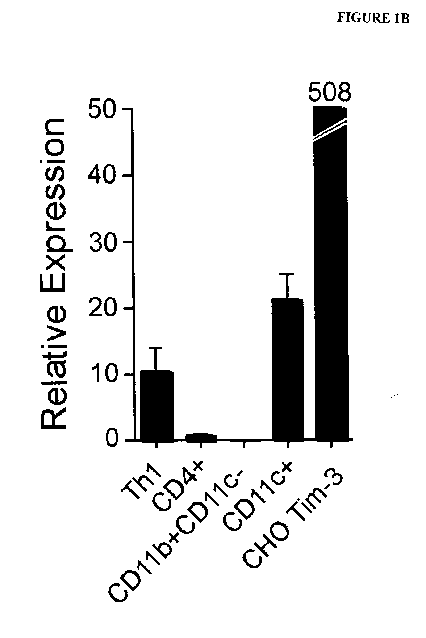
[0313]Pure populations of both macrophages and dendritic cells were isolated by cell sorting, and examined for TIM-3 mRNA by quantitative RT-PCR. mRNA from ex vivo sorted CD4+ T cells, an activated Th1 T cell clone and CHO TIM-3 transfectants were used as controls. In agreement with the flow cytometry data, TIM-3 mRNA was present at high levels in dendritic cells and absent in macrophages (FIG. 1B).


PUM

PropertyMeasurementUnit
Fractionaaaaaaaaaa
Immunogenicityaaaaaaaaaa
Levelaaaaaaaaaa

Abstract

The invention provides novel methods of treating neurological disorders, including neurodegenerative disorders such as MS. The invention also provides novel methods of treating cancers, including glial tumors such as glioblastoma multiforme. The invention further provides vaccines and related uses.

Description

STATEMENT REGARDING FEDERALLY-SPONSORED RESEARCH OR DEVELOPMENT[0001]The invention described herein was supported, in whole or in part, by the National Institute of Health Grant Nos P01NS038087 and NS045937. The United States government has certain rights in the invention.BACKGROUND OF THE INVENTION[0002]Multiple sclerosis (“MS”) affects approximately 1 out of 1,600 people in the United States and is a common cause of persistent disability in young adults. MS involves repeated episodes of inflammation of central nervous system tissue in any area of the brain and spinal cord. The inflammation destroys the myelin sheath covering the nerve cells in the affected area. This leaves multiple areas of scar tissue (sclerosis) along the covering of the nerve cells. Sclerosis slows or blocks the transmission of nerve impulses in that area, resulting in the development of the symptoms of MS.[0003]The location of the inflammation varies from person to person and from episode to episode causing a...

Claims


Application Information

Patent Timeline
IPC IPC(8): A61K39/395A61K38/17A61K48/00A61P37/00A61K39/00A61P35/00
CPCA01K2227/105A01K2267/0387C07K2316/95C07K16/28C07K14/705A61P3/02A61P13/10A61P15/10A61P21/00A61P25/00A61P25/28A61P27/16A61P35/00A61P37/00A61P37/04C07K2317/75
Inventor ANDERSON, DAVID E.ANDERSON, ANA C.KUCHROO, VIJAY K.HAFLER, DAVID A.
Owner THE BRIGHAM & WOMEN S HOSPITAL INC
Why Eureka
Try Eureka