
Method of detecting cellular immunity and application thereof to drugs

a technology of cellular immunity and antigens, applied in the field of immunotherapy, can solve the problems of large number, difficult to determine whether a particular spot is positive or not, complex process, etc., and achieve the effect of increasing the expression of cell surface antigens and expressing stably

Inactive Publication Date: 2006-02-16
GREEN PEPTIDE CO LTD +1
View PDF1 Cites 20 Cited by
  • Summary
  • Abstract
  • Description
  • Claims
  • Application Information

AI Technical Summary

Benefits of technology

[0065] The frequency of peptide stimulation has traditionally been once a week, over a few weeks (usually about three), and it has been thought that there is no need to do more. However, it is shown herein that frequent stimulation, for example, several times at intervals of two to five days, preferably induces antigen-specific T cells. Therefore, the “frequent” of frequent stimulation herein signifies multiple stimulations at a frequency of more than twice a week or at intervals of two to five days. It has been shown that induction of antigen-specific T cells by frequent stimulation enables detection in a relatively short period of time, about 13 days, compared to traditional techniques. Therefore, “multiple times” herein signifies stimulation in a certain time period of less than 15 days, preferably less than 13 days. Stimulation for more than 15 days does not significantly improve the effect of the invention, which diminishes the need for it from the standpoint of medical treatment. If used multiple times, for example stimulated at an interval of 3 days, the total number of stimulations is four, including the initial stimulation. Multiple stimulations can be performed by exchanging a portion of the culture supernatant with the same amount of fresh lymphocyte medium containing the antigen.
[0066] The detection of the antigen-specific cells can be performed by various methods. One such method is, to react the post-stimulated antigen-specific T cells to the appropriate targeted cells, thereby quantifying the cytokine produced by the T cells, preferably IFN-γ. However, the invention is not limited to this method and can be applied to any method that can detect antigen-specific T cells unless the method adversely affects the detection process.
[0067] To prepare the targeted cells, first, the HLA gene which matched the HLA of the antigen-specific T cells was introduced to various cell lines not expressing those antigens, and was expressed stably. In other words, cells, which do not express antigens, in which the cDNA expressing plasmid of the HLA gene which is matched with HLA of antigen-specific T cells, can be employed as target cells. Various cell lines expressing HLA matched with antigen-specific T cells but not expressing antigens can be also used for the target cells.
[0068] Using the antigen or antigen peptide used for the stimulation of peripheral blood mononuclear cells, the prepared cells are sensitized as antigen-presenting cells. Peptides other than the antigen or antigen peptide used for the stimulation of peripheral blood mononuclear cells, sensitize the prepared cells which are used as target cells as a control.
[0069] Cytokine levels, or better still INF-γ levels, can be quantified after the reaction with cultured peripheral blood mononuclear cells using a method such as ELISA or FLISA, and the production of desired antigen-specific T cells can be assessed by comparison with the control.
[0070] The advantage of quantification of cytokines, especially INF-γ is that INF-γ is produced by various subtypes of lymphocytes, such as CD4+, CD8+, NK, LAK cells, and the quantification has been widely used to monitor immune reaction in many clinical studies of cancer vaccines because the INF-γ possesses a variety of biological characteristics, for example, immunomodulation activity, antivirus activity, increased expression of cell surface antigens like HAL molecules, and is secreted after stimulation with antigens.

Problems solved by technology

In this method, the process is complicated because the lymphocytes used as effector cells are sensitized with antigen peptide multiple times, once a week in order to improve the detection sensitivity of the antigen-specific T cells.
In this method, since the noise is high, determining whether a particular spot is positive or not may be difficult.
However, these methods employ stimulation by antigen presenting cells (APCs) and induction of antigen-specific CTL from peripheral-blood mononuclear cells (PBMCs) in a cancer patient, and they require a considerable number of peripheral-blood mononuclear cells as feeder cells in order to study the reactivity to a cluster of peptides.
As set forth above, each traditional method of detecting antigen-specific cells has some problems associated with it, such as the amount of blood to be collected, the complexity of the procedure and detection sensitivity.

Method used


Image

  • Method of detecting cellular immunity and application thereof to drugs
  • Method of detecting cellular immunity and application thereof to drugs
  • Method of detecting cellular immunity and application thereof to drugs

Examples


example 1

(Detection of Antigen Peptide-specific T Cells Using an EB Virus-derived Antigen Peptide)

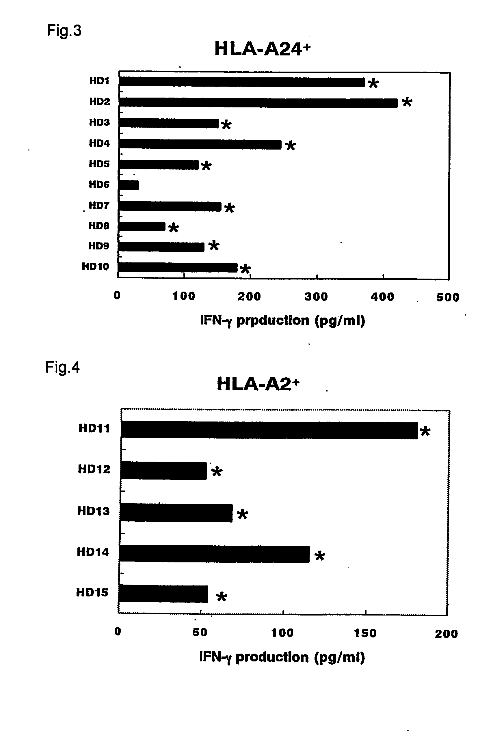
[0090] Most healthy people retain EB virus (EBV)-specific T cell precursors relatively frequently from a past EB virus infection. Therefore, we tried to detect the EB virus-specific T cell from peripheral blood mononuclear cells (PBMCs) of healthy donors with a history of EB virus infection with positive blood anti-EB virus antibody.

[0091] The PBMCs were isolated from the peripheral blood of HLA-A24 positive healthy people by the specific gravity centrifugal method using Ficoll Conray solution. The PBMCs were cultured in a medium wherein to 45% RPMI-1640, 45% AIM-V (GIBCO BRL Inc.), and 10% FCS, 100 IU / ml of interleukin-2 (IL-2), 0.1 mM MEM nonessential amino acid solution (GIBCO BRL Inc., hereinafter called “NEAA”) were added (hereinafter called lymphocyte medium). PBMCs were suspended in the lymphocyte medium at 5×10⁵ cells / ml and inoculated in a 96-well plate having U-shaped bottoms at 200...

example 2

(Analysis of the Frequency of EB Virus Derived Antigen Peptide-Specific T cells)

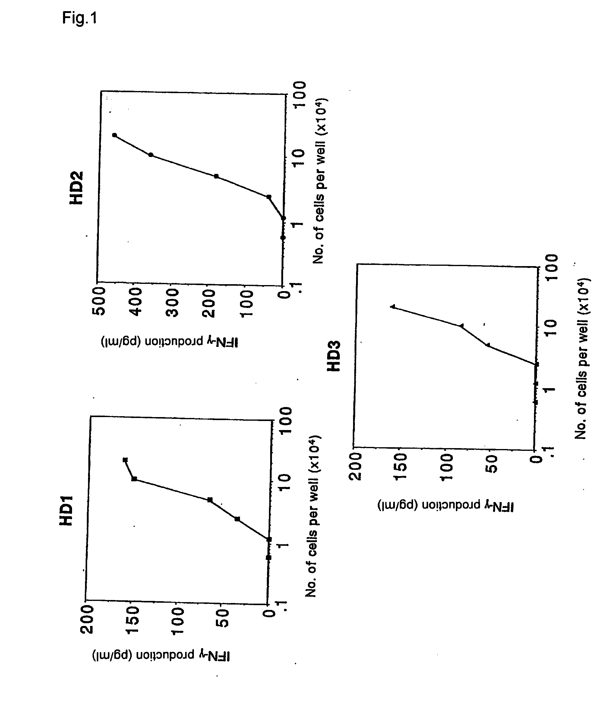
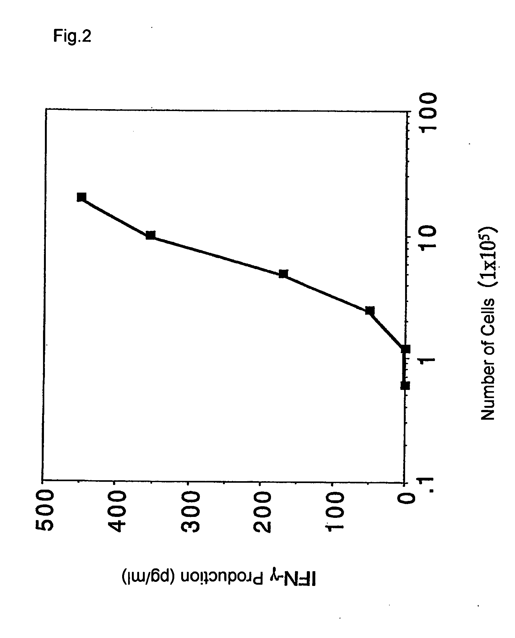
[0093] (1) According to the procedure in example 1, the increase in frequency of EB virus derived HLA-A24 restricted antigen peptide-specific T cells was confirmed by limiting dilution analysis.

[0094] The PBMCs were prepared from the same healthy donor as Example 1 by the same procedure, and the following measurement of frequency of antigen specific T cells was performed with or without stimulating a portion of the PBMCs using an antigen peptide. Another portion of PBMCs were added to a 96-well plate having wells with a U-shaped bottom at a concentration of 1×10⁵ cells / well in a method similar to that in Example 1. These were cultured while being stimulated using EB virus derived HLA-A24 restricted antigen peptides four times in total every three days. The cultures were subsequently measured for the frequency of the antigen-specific T cells as described below.

[0095] The measurement of the frequency o...

example 3

(Peptide Stimulated CTLs and Their Specificity)

[0099] To isolate PBMCs, blood samples collected from healthy donors and patients were heparinized and PBMCs were obtained by density gradient electrophoresis. All donors of the blood samples were serologically HIV negative and HTLV-1 negative.

[0100] Various densities of PBMCs were cultured by adding them to 96-well microplates having wells with a U-shaped or flat bottom with 200 μl of medium containing 10 μM of an antigen peptide. The medium used consisted of a composition of 45% RPMI-1640, 45% AIM-V, 10% FCS, 100 IU / ml of IL-2, and 0.1 μM of NEAA.

[0101] The antigen peptides used were the following: [0102] EB virus derived peptide comprising HLA-A24 binding motif (sequence of amino acids: TYGPVFMCL) (SEQ ID No: 31); [0103] EB virus derived peptide comprising HLA-A2 binding motif (sequence of amino acids: GICTLVAML) (SEQ ID No: 32); [0104] Influenza virus (Flu) derived peptide comprising HLA-A24 binding motif (sequence of amino acid...


PUM

PropertyMeasurementUnit
time periodaaaaaaaaaa
focal depthaaaaaaaaaa

Abstract

It is intended to provide a convenient immunity monitoring system whereby T cell frequencies specific to a plural number of antigen peptides can be assayed by using a relatively small amount of blood. Peripheral monocytes are collected and frequently stimulated with an antigen without directly using any antigen presenting cells. Then T cells specific to the antigen in the thus stimulated peripheral monocytes are detected to thereby detect antigen-specific T cells. thus, diseases such as cancer can be prevented or treated with the use of a peptide having such a function, in particular, cancer tumor-rejection antigen peptide.

Description

FIELD OF THE INVENTION [0001] This invention relates to the field of immunotherapy and to a method of detecting cellular immunity and a procedure for use thereof. More specifically, the invention relates to a method of detecting antigen-specific T cells and a procedure for use thereof. BACKGROUND OF THE INVENTION [0002] Thanks to the latest developments in the field of cellular immunology and molecular biology, many epitope peptides recognized by cytotoxic T lymphocytes (CTLs), which react to cancerous or virus-infected cells, have been identified. As a result of such developments, we have found new peptide-based immunotherapies for cancers, virus infections, and various autoimmune diseases. T cells associated with cellular immunity, especially antigen-specific T cells, have an important role in the elimination of cancerous or virus-infected cells by a body. Antigen-specific T cells are also known to be involved in autoimmune diseases such as multiple sclerosis and rheumatoid arthri...

Claims


Application Information

Patent Timeline
Patent Type & Authority Applications(United States)
IPC IPC(8): G01N33/574G01N33/50
CPCA61K39/0011G01N33/505G01N33/5011A61K39/35A61K39/001178
Inventor ITOH, KYOGOHIDA, NAOYA
Owner GREEN PEPTIDE CO LTD
Learn More