Bio-barcode based detection of target analytes

a technology of biobarcode and target analytes, applied in the field of biobarcode based detection of target analytes, can solve the problems of increasing assay time, difficult, expensive, time-consuming, etc., and simultaneously detecting several protein structures using the aforementioned related protocols

Inactive Publication Date: 2006-02-23
NORTHWESTERN UNIV
View PDF89 Cites 79 Cited by
  • Summary
  • Abstract
  • Description
  • Claims
  • Application Information

AI Technical Summary

Benefits of technology

[0053] As discussed herein, the assay methods of the invention are ultrasensitive and can be performed in less than 60 minutes without the need for enzymatic amplification or a scanometric detection scheme. By eliminating both PCR and scanometric method without losing sensitivity, the methods of the invention overcome some of the drawbacks that both of these conventional methods, such as long assay times and assay complexity. Moreover, non-scientists such as nurses, medical doctors, and soldiers should be able to learn how to use the methods of the invention after simple training due to its simplicity. The methods of the invention can be easily multiplexed by using different fluorophores for different target proteins and total assay time can be even more decreased by optimizing probe concentrations and reaction conditions. In addition, the methods of the invention can be used to detect any target that can be detected in a sandwich assay, including proteins, DNA, RNA, small molecules, and metal ions. Furthermore, other types of reporter groups can be attached to the barcodes allowing for methods other than fluorescence-detection to identify the barcodes and amplified detection signal. These include, but are limited to, redox-active groups, groups with electrical signatures, radioactive groups, catalytic groups, groups with distinct absorption characteristics, and groups with Raman signatures. A reporter group can be anything with a distinct and measurable chemical or physical signature.

Problems solved by technology

However, it is difficult, expensive, and time-consuming to simultaneously detect several protein structures under assay conditions using the aforementioned related protocols.
Although theses approaches are notable advances in protein detection, they have several drawbacks: 1) limited sensitivity because of a low ratio of DNA identification sequence to detection antibody; 2) slow target binding kinetics due to the heterogeneous nature of the target capture procedure, which increases assay time and decreases assay sensitivity (Step 3 in FIG. 5); 3) complex conjugation chemistries that are required to chemically link the antibody and DNA-markers (Step 4 in FIG. 5); and 4) require a PCR amplification step45.

Method used


Image

  • Bio-barcode based detection of target analytes
  • Bio-barcode based detection of target analytes
  • Bio-barcode based detection of target analytes

Examples


example 1

Preparation of Oligonucleotide-Modified Gold Nanoparticles

A. Preparation of Gold Nanoparticles

[0476] Oligonucleotide-modified 13 nm Au particles were prepared by literature methods (˜110 oligonucleotides / particle)18-20. Gold colloids (13 nm diameter) were prepared by reduction of HAuCl4 with citrate as described in Frens, Nature Phys. Sci., 241, 20 (1973) and Grabar, Anal. Chem., 67, 735 (1995). Briefly, all glassware was cleaned in aqua regia (3 parts HCl, 1 part HNO3), rinsed with Nanopure H2O, then oven dried prior to use. HAuCl4 and sodium citrate were purchased from Aldrich Chemical Company. An aqueous solution of HAuCl4 (1 mM, 500 mL) was brought to a reflux while stirring, and then 50 mL of a 38.8 mM trisodium citrate solution was added quickly, which resulted in a change in solution color from pale yellow to deep red. After the color change, the solution was refluxed for an additional fifteen minutes, allowed to cool to room temperature, and subsequently filtered through a...

example 2

Preparation of Hapten-Modified Oligonucleotides

[0483] Hapten-modified oligonucleotides were prepared with a biotin-triethylene glycol phosphoramidite for A1 and 2,4-dinitrophenyl-triethylene glycol phosphoramidite for B1 (Glen research) using standard solid-phase DNA synthesis procedures.21

[0484] Biotin modified oligonucleotides were prepared using the following protocol: A CPG-bound, detritylated oligonucleotide was synthesized on an automated DNA synthesizer (Expedite) using standard procedures21. The CPG-cartridge was then removed and disposable syringes were attached to the ends. 200 uL of a solution containing 20 umole of biotin-triethylene glycol phosphoramidite in dry acetonitrile was then mixed with 200 uL of standard “tetrazole activator solution” and, via one of the syringes, introduced into the cartridge containing the oligonucleotide-CPG. The solution then was slowly pumped back and forth through the cartridge for 10 minutes and then ejected followed by washing with dr...

example 3

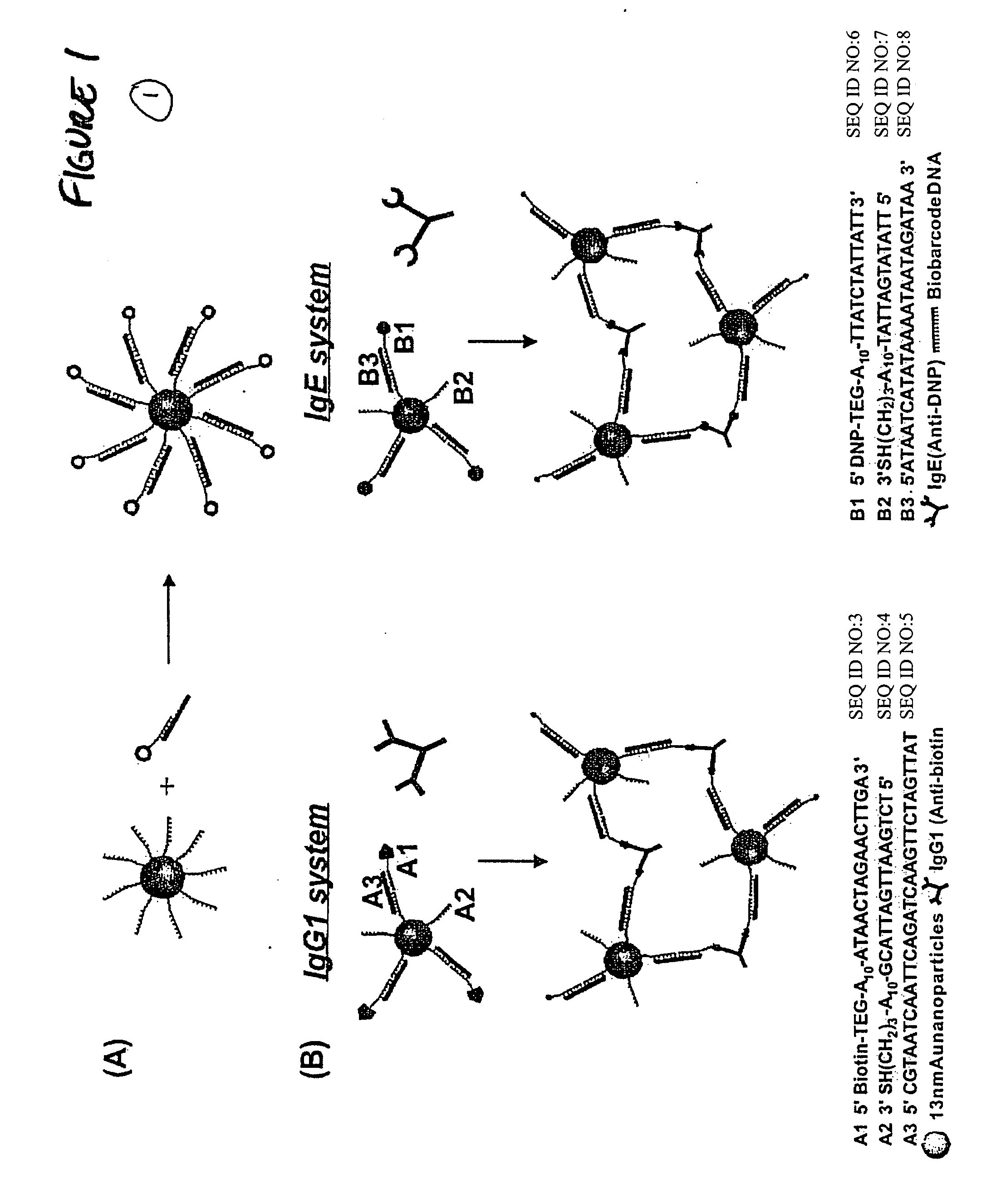
Assay Using Nanoparticle Complex Probes

[0485] The Oligonucleotide-modified 13 nm gold particles were prepared as described in Example 1. Hapten-modified oligonucleotides were prepared as described in Example 2 with a biotin-triethylene glycol phosphoramidite for A1 and 2,4-dinitrophenyl-triethylene glycol phosphoramidite for B1 (Glen research) using standard solid-phase DNA synthesis procedures.21 The PBS buffer solution used in this research consists of 0.3 M NaCl and 10 mM phosphate buffer (pH 7). IgE and IgG1 were purchased from Sigma Aldrich (Milwaukee, Wis.) and dissolved in 0.3 M PBS buffer with 0.05% Tween 20 (final concentration: 4.3×10⁻⁸ b / μl) and background proteins (10 ug / ml of lysozyme, 1% bovine serum albumin, and 5.3 ug / ml of anti-digoxin; 10 uL of each) prior to use.

[0486] To prepare the probes, the oligonucleotide modified particles (13 nM, 300 μL) were hybridized with hapten-modified complementary oligonucleotides (10 μL of 10 μM) and biobarcode DNA (10 μL of 10 μ...


PUM

PropertyMeasurementUnit
diameteraaaaaaaaaa
volumeaaaaaaaaaa
diameteraaaaaaaaaa

Abstract

The present invention relates to screening methods, compositions, and kits for detecting for the presence or absence of one or more target analytes, e.g. biomolecules, in a sample. In particular, the present invention relates to a method that utilizes reporter oligonucleotides as biochemical barcodes for detecting multiple protein structures or other target analytes in a solution.

Description

CROSS-REFERENCE [0001] This application is a continuation-in-part of U.S. patent application Ser. No. 10 / 877,750, filed Jun. 25, 2004 and International application PCT / US04 / 020493, filed Jun. 25, 2004, both of which are incorporated by reference in their entirety. This application claims the benefit of provisional application Nos. 60 / 570,723, filed May 12, 2004; 60 / 585,294, filed Jul. 1, 2004; and ______ (Atty Docket No. 05-192) filed Jan. 19, 2005. This application also claims the benefit of provisional application Nos. 60 / 506,708, filed Sep. 25, 2003; 60 / 482,979, filed Jun. 27, 2003; 60 / 496,893, filed Aug. 21, 2003; 60 / 515,243, filed Oct. 28, 2003; 60 / 530,797, filed Dec. 18, 2003 and is a continuation-in-part of U.S. patent application Ser. No. 10 / 108,211, filed Mar. 27, 2002, which in turn claims the benefit of U.S. Provisional application Nos. 60 / 192,699, filed Mar. 28, 2000; and 60 / 350,560, filed Nov. 13, 2001, which are incorporated by reference in their entirety, and which is...

Claims


Application Information

Patent Timeline
Patent Type & Authority Applications(United States)
IPC IPC(8): C12Q1/68
CPCC12Q1/6816G01N33/54333C12Q2565/501C12Q2565/113C12Q2563/179C12Q2563/155C12Q2563/143C12Q2563/149
Inventor MIRKIN, CHADNAM, JWA-MINOH, BYUNG-KEUNTHAXTON, C. SHADGEORGANOPOULOU, DIMITRA
Owner NORTHWESTERN UNIV
Why Eureka
Try Eureka