

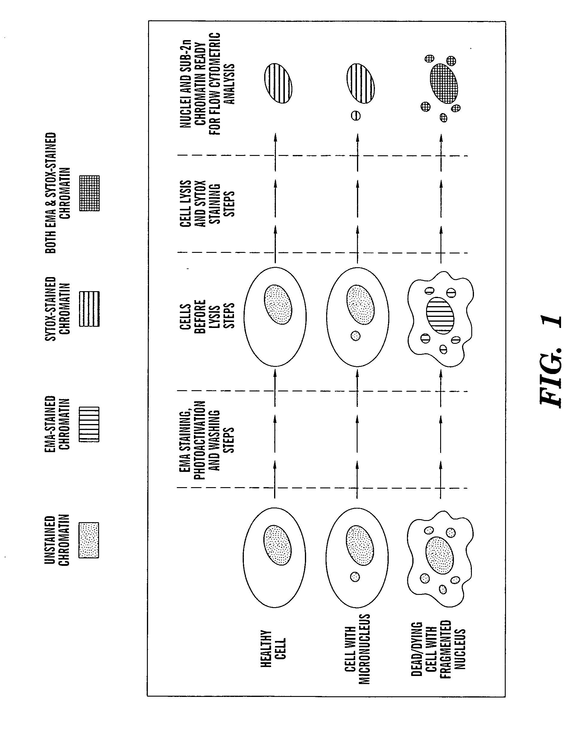
Method for enumerating mammalian cell micronuclei with an emphasis on differentially staining micronuclei and the chromatin of dead and dying cells
a technology of mammalian cell micronuclei and chromatin, applied in the field of enumerating mammalian cell micronuclei with, can solve the problems of insufficient toxicity evaluation of many natural and industrially manufactured compounds and formulations, laborious traditional toxicity evaluation, and inability to demonstrate acceptable toxicity to critical organs, etc., to achieve accurate and reliable micronuclei measurements, cost savings, and fast, reliable and accurate results
- Summary
- Abstract
- Description
- Claims
- Application Information
AI Technical Summary
Benefits of technology
Problems solved by technology
Method used
Image

Examples
example 1
Heat Shock Treatment
[0079] L5 1 78Y cells were exposed to an elevated temperature (“heat shock”) to evaluate the stage of apoptosis at which EMA staining occurs. Other more commonly used labeling techniques were studied to provide a frame of reference. Representative time-course data are presented in FIG. 3. As expected, cell surface expression of phosphatidyl serine, as demonstrated by Annexin V-labeling, occurred most rapidly in response to heat treatment. In contrast, exclusion of the charged nucleic acid dye propidium iodide was only modestly affected over this time-course. Relative to annexin and propidium iodide, an intermediate rate of EMA-positive labeling was observed. In fact, EMA performed quite similarly to YO-PRO-1, a nucleic acid dye that has been described as a relatively early-stage apoptosis marker (Idziorek et al., “YOPRO-1 Permits Cytofluorometric Analysis of Programmed Cell Death (Apoptosis) Without Interfering with Cell Viability,”J. Immunol. Methods 185:249-25...
example 2
Genotoxicants
[0082] Each of the six genotoxicants studied was observed to cause a dose-related increase in micronucleus frequency (FIGS. 5A-G). As demonstrated by the low degree of variation among replicate flow cytometric samples, reproducible % micronuclei values were observed for the automated scoring process for both vehicle and genotoxicant-treated cultures. Furthermore, the correspondence between flow cytometric- and microscopy-based values was high (rs values ranged from 0.7 to 1.0; see Table 2).
TABLE 2Overview of Micronucleus Test Results (L5178Y Cells)HighestTreatment / Conc.MNFCM / MicroscopyCellRecoveryTestedInductionSpearmanCycleChemical(hrs)(μg / ml)(≧3-fold)CorrelationEffect*Methyl4 / 2030Yes1.000+++methanesulfonateHydroxyurea4 / 2018Yes0.9429+++Etoposide4 / 200.1Yes1.000++Cyclophosphamide4 / 206Yes1.000+monohydrateBenzo[a]pyrene4 / 204Yes0.9370−Vinblastine sulfate4 / 200.3Yes0.7000+++Vinblastine sulfate24 / 0 0.00075Yes1.000+++Tributyltin4 / 200.15No0.9487−methoxideTributyltin24 / 0 0.06N...
example 3
Non-Genotoxicants
[0085] As shown in FIG. 5H, sucrose was neither cytotoxic nor genotoxic up to 5 mg / ml in the short-term exposure scenario. Based on expert working group recommendations, negative short-term results were followed by a continuous-treatment exposure (Kirsch-Volders et al., “Report from the In Vitro Micronucleus Assay Working Group,”Mutat. Res. 540:153-163 (2003), which is hereby incorporated by reference in its entirety). Again, micronucleus-induction was not evident (see FIG. 51). Correlation to microscopy is poorly depicted by the rs values that appear in Table 2. This may be explained by the extreme sensitivity of this statistic when % micronuclei frequencies are fluctuating in the range of baseline values.
[0086] The apoptosis-inducing agents tributyltin methoxide and dexamethasone were also investigated in both short-term and continuous exposure scenarios. Unlike sucrose, these experiments included concentrations which significantly affected cell survival (FIGS. ...
PUM
| Property | Measurement | Unit |
|---|---|---|
| Time | aaaaa | aaaaa |
| Time | aaaaa | aaaaa |
| Time | aaaaa | aaaaa |
Abstract
Description
Claims
Application Information