
Detection of microorganisms with holographic sensors

a technology of holographic sensor and microorganism, applied in the field of cell detection, can solve the problems of high cost, sensitive to environmental contamination, and inability to accurately identify a microbial pathogen, and achieve the effect of reducing the number of experiments

Inactive Publication Date: 2006-03-16
SMART HOLOGRAMS
View PDF6 Cites 3 Cited by
  • Summary
  • Abstract
  • Description
  • Claims
  • Application Information

AI Technical Summary

Benefits of technology

"The invention is a method and device for detecting cells using an optical sensor. The cell is immobilised using an antibody and a growth medium is introduced. The sensor detects a product of the cell's growth and a change in its optical characteristic is detected. The device is simple to operate and compatible with standard laboratory techniques. It can be integrated with PCR technology for full integration with laboratory-based diagnostics. The technical effect of the invention is rapid, accurate identification of target organisms with the specificity of ELISA technology, even at sub-infectious concentrations, making it useful in a wide range of conditions."

Problems solved by technology

Whilst there are a number of competing technologies available to aid in this process, such as ELISA and PCR, the definitive identification of a microbial pathogen is still a time-consuming, laboratory-based procedure.
Unfortunately, this technology is sensitive to environmental contamination, meaning that sample pre-treatment is necessary in many instances.
This technology is also expensive and requires highly trained personnel.
Neither of these methods is readily compatible with conventional microbiology techniques.
While they may be used in some circumstances to determine the identity of a microbe in a large or pure sample, they do not readily lend themselves to direct comparison with laboratory assays in which cells are cultured and identified using classical microbiological methodologies.
Nor do they provide a means for capturing viable cells for definitive identification.

Method used


Image

  • Detection of microorganisms with holographic sensors
  • Detection of microorganisms with holographic sensors
  • Detection of microorganisms with holographic sensors

Examples


example 1

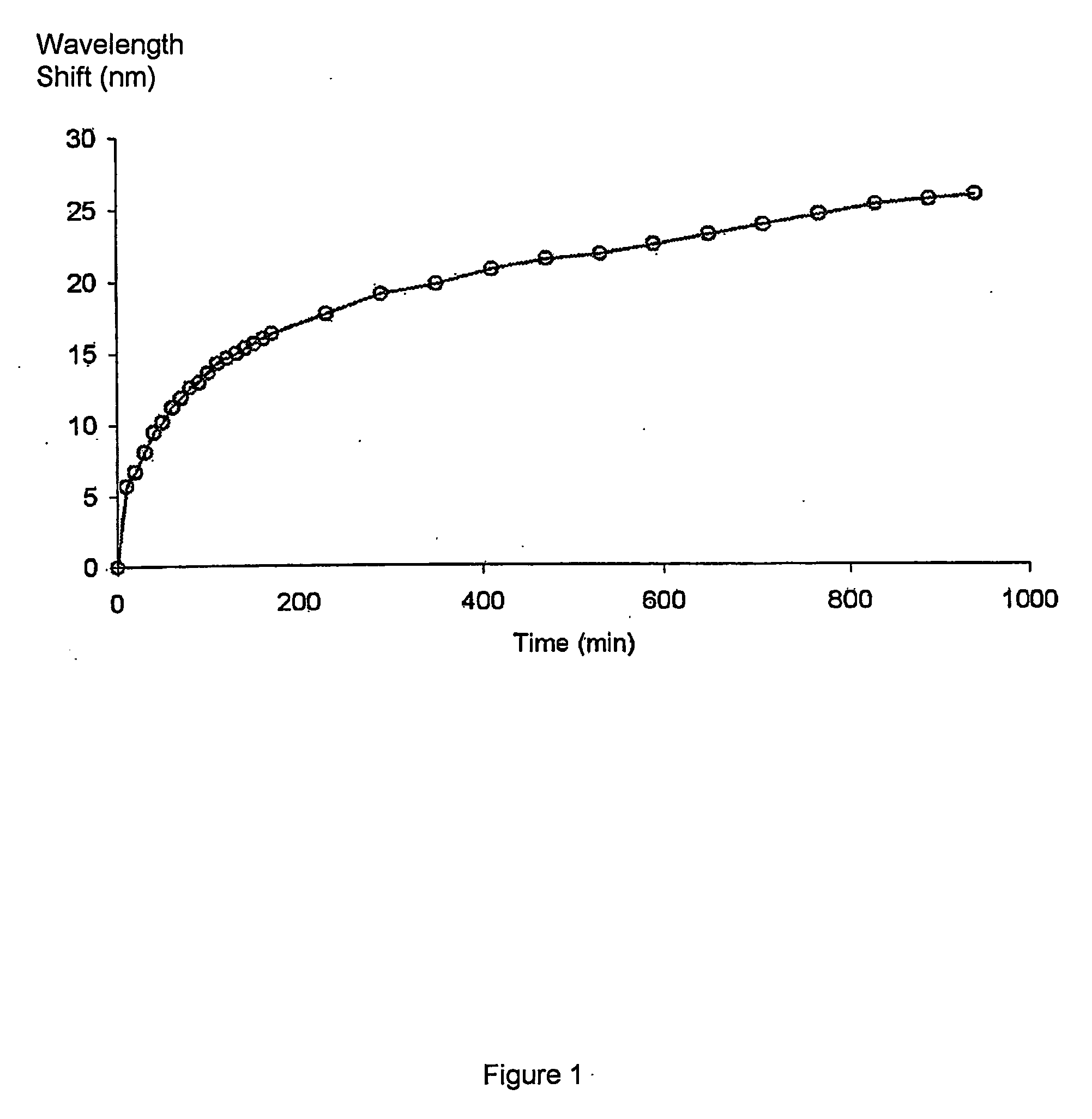
[0025]Bacillus subtilis was detected in microbial culture. A metabolic product of the bacterium is protease, which degrades a gelatin-based holographic sensor. As the gelatin support medium degrades, it becomes increasingly spongy and expands.

[0026] Mid-exponential phase culture (in nutrient broth) was inoculated into a cuvette containing the hologram, and a reflection spectrometer used to measure the peak wavelength at 10 minute intervals over 15 hours at 30° C. A positive result for protease was shown by the peak wavelength undergoing a red-shift. FIG. 1 shows the red-shift of the peak wavelength of reflection over the 15 hour period.

example 2

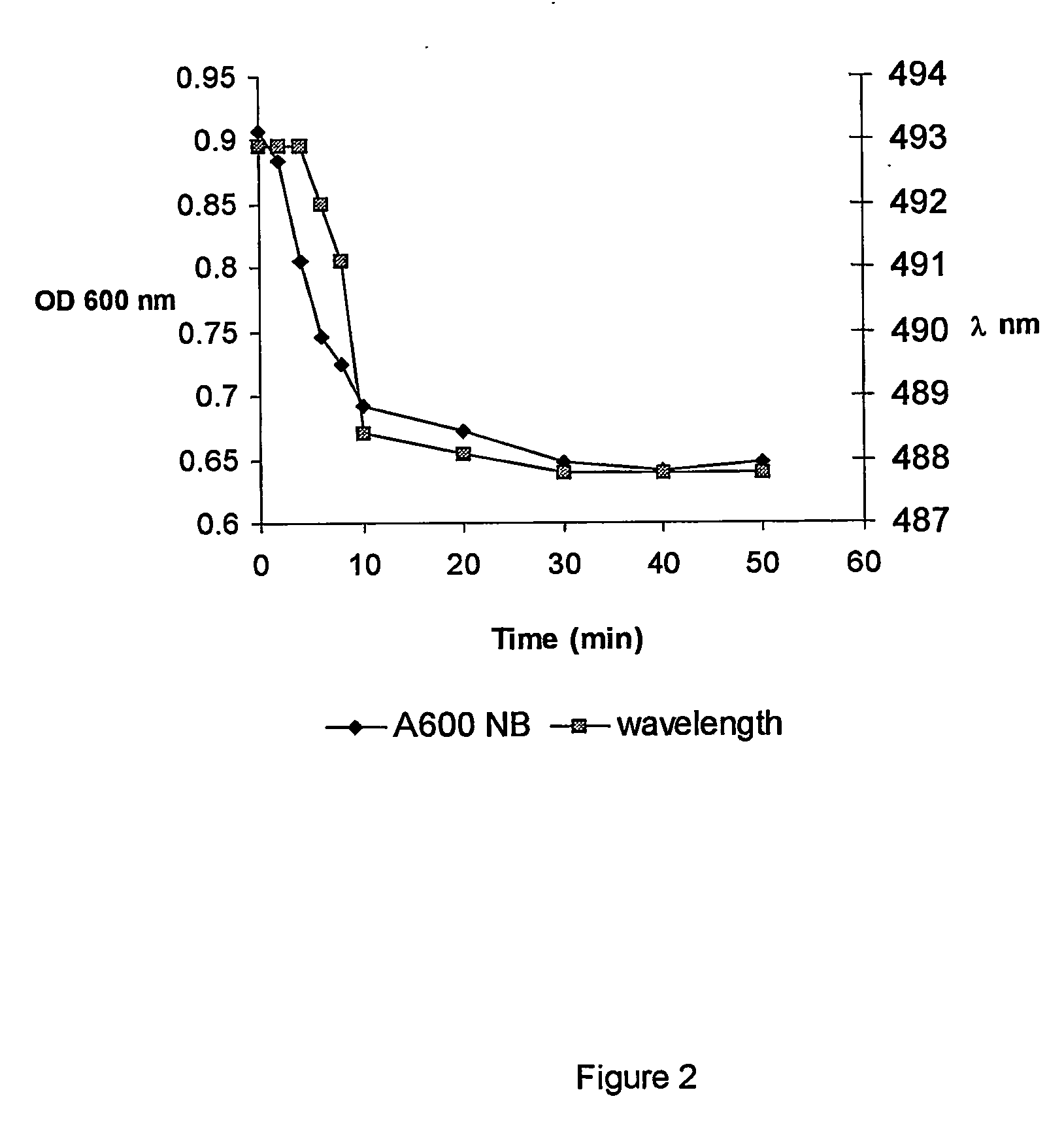
[0027]Bacillus megaterium was detected in microbial culture. During germination, the bacterium releases Ca2+ (bound to dipicolinic acid). Ca2+ binds to a polyHEMA-MIDA holographic support medium, inducing a concomitant contraction of the polymer and a shift in replay wavelength.

[0028] A holographic sensor compound of 10 and 12 mole % MIDA in polyHEMA was equilibrated in nutrient broth. Bacillus megaterium spores were then added at a concentration of approximately 108 spores / ml. A reflection spectrometer was used to measure the peak wavelength at 1 minute intervals for 50 minutes at 25° C. Any change in the optical density of the sensor was also detected, a change in optical density being indicative of germination. Changes in the optical density of the germination matrix were also detected.

[0029]FIG. 2 is a graph of the germination response, showing the optical density (OD) and wavelength readings. The decreases in both OD and A are indicative of Ca2+-induced binding to of the holo...

example 3

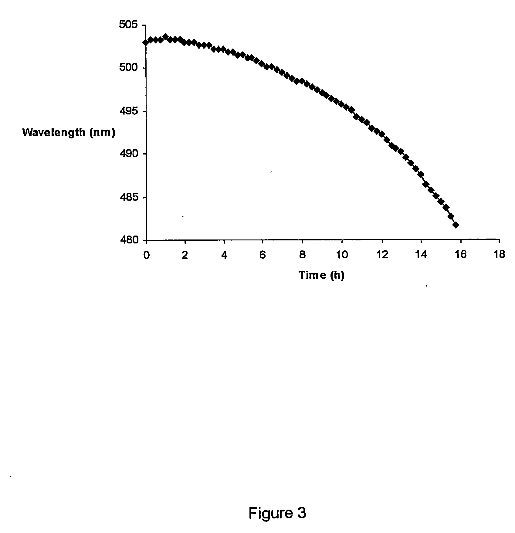
[0030] Vegetative Bacillus megaterium was detected using a starch / acrylamide holographic sensor. The Bacillus genus is characterised by relatively high amylase production during growth; amylase degrades a starch-based holographic support medium.

[0031] A section of the sensor was equilibrated with 1800 μl of nutrient both at 30° C. 200 μl of vegetative Bacillus megaterium (cultured overnight) was then added (the cells were centrifuged and resuspended in fresh medium prior to addition to the cuvette, to remove any residual amylase). The peak wavelength of reflection of the sensor was recorded every 15 minutes for approximately 16 hours.

[0032] The results are shown in FIG. 3. Initially, the shift in wavelength was relatively small; however, the shift was more pronounced with time. This lag may be due to the presence of residual glucose in the holographic support medium.


PUM

PropertyMeasurementUnit
optical characteristicaaaaaaaaaa
magnetic fieldaaaaaaaaaa
volumeaaaaaaaaaa

Abstract

A method for the detection of a cell comprises immobilizing the cell in a device also containing a sensor, and introducing a growth medium, wherein the sensor is sensitive to a product of the cell's growth; and detecting any change in an optical characteristic of the sensor. A device suitable for use in the invention comprises a chamber including a sensor, inlets for sample and for a growth medium, and means for immobilizing an antibody in the chamber or elsewhere in the device that provides a fluidic link with the sensor.

Description

FIELD OF THE INVENTION [0001] This invention relates to the detection of cells, e.g. using a holographic sensor. BACKGROUND TO THE INVENTION [0002] Rapid identification of cells, in particular pathogenic cells, is of vital importance in diagnostics and biodefence. Whilst there are a number of competing technologies available to aid in this process, such as ELISA and PCR, the definitive identification of a microbial pathogen is still a time-consuming, laboratory-based procedure. [0003] ELISA kits for the detection of agents such as Bacillus anthracis are available. These kits are highly specific to the target organism, showing no cross-reaction with closely related Bacillus species. They are, however, somewhat insensitive, requiring in the order of 10,000 cells, in order to avoid false negatives; this quantity of cells is somewhat more than a human infective dose of a microbe such as Bacillus anthracis. [0004] PCR technology provides a fast, accurate and rapid means for determining t...

Claims


Application Information

Patent Timeline
Patent Type & Authority Applications(United States)
IPC IPC(8): G01N33/554C12Q1/04C12M1/34G01N33/569G01N21/27G01N21/77
CPCG01N21/77C12Q1/04
Inventor LOWE, CHRISTOPHER ROBINDAVIDSON, COLIN ALEXANDER BENNETT
Owner SMART HOLOGRAMS
Learn More