Telomelysin/GFP-expressing recombinant virus

a virus and telomelysin technology, applied in the field of telomely, can solve the problems of insufficient development of the infected living body detection system during surgical operation, and no research has been known to date in which the living body is infected with a virus

Active Publication Date: 2006-03-30
ONCOLYS BIOPHARMA
View PDF0 Cites 27 Cited by
  • Summary
  • Abstract
  • Description
  • Claims
  • Application Information

AI Technical Summary

Benefits of technology

[0006] As a result of intensive and extensive researches toward the solution of the above problems, the present inventors have found that it is possible to detect cancer cells with extremely high sensitivity and even in vivo, by integrating a gene encoding a fluorescence la...

Problems solved by technology

However, in situ cancer detection system during surgical operation has not yet been developed because of the difficulty in targeting cancer cells.
Further, no research...

Method used


Image

  • Telomelysin/GFP-expressing recombinant virus
  • Telomelysin/GFP-expressing recombinant virus
  • Telomelysin/GFP-expressing recombinant virus

Examples


example 1

Visualization of Cancer Cells by in Vitro Co-Infection

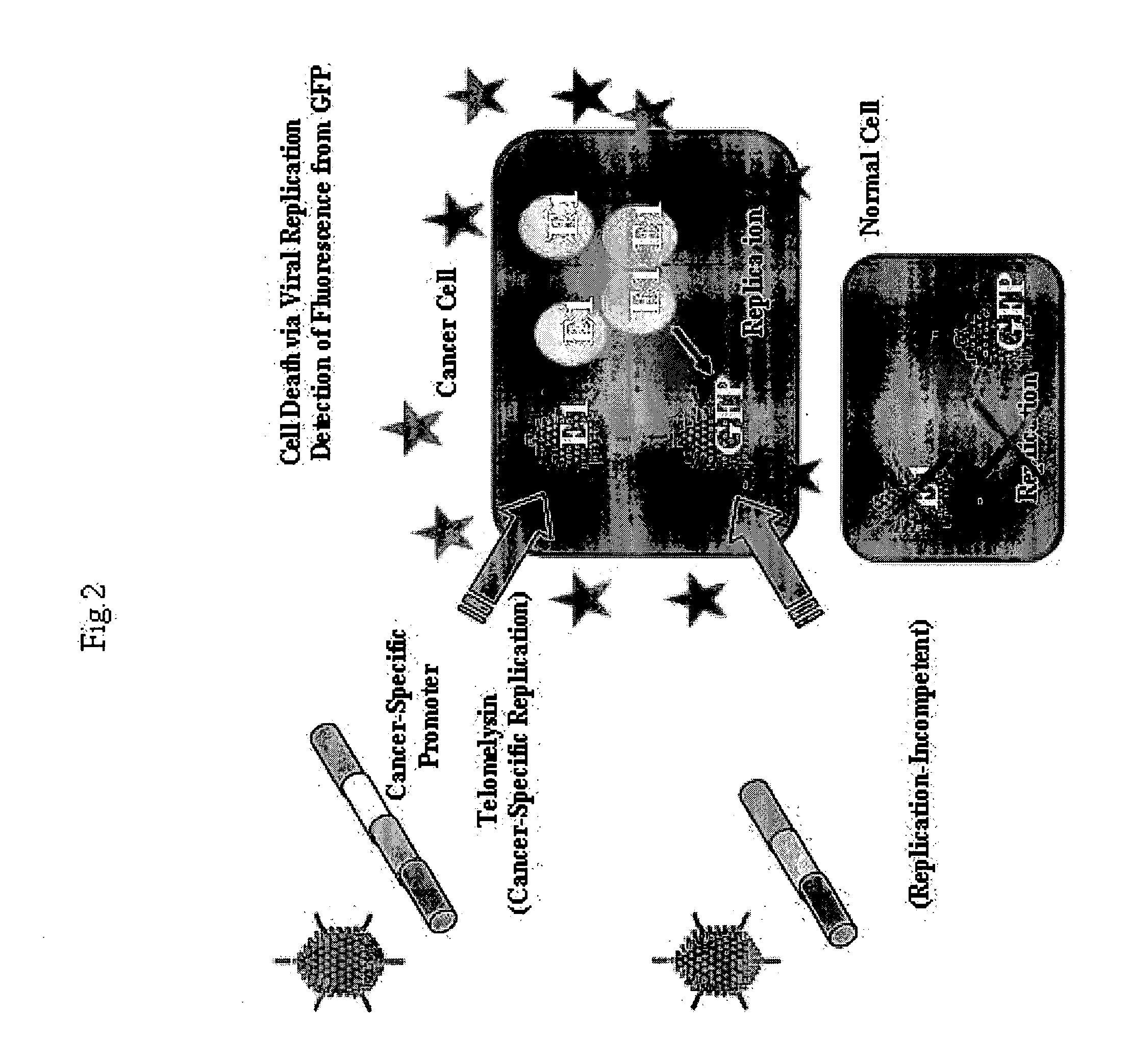
[0104] This Example preliminary examined whether or not fluorescence will be emitted in vitro when cancer cells are co-infected with virus Telomelysin comprising the replication cassette and non-replicating virus Ad-GFP comprising the labeling cassette.

[0105] Human large colon cancer cell SW620 and human lung cancer cells A549 and H1299 were infected with 0.1 MOI (multiplicity of infection) of Ad-GFP (FIG. 2).

[0106] As a result, tendency to green color was hardly recognized when human large colon cancer cell SW620 and human lung cancer cells A549 and H1299 were infected with 0.1 MOI of Ad-GFP. However, when 1 MOI of TRAD was used jointly, fluorescence could be detected only in cancer cells, and no fluorescence was detected in normal cells such as human fibroblast cells W138 and NHLF and human umbilical vascular endothelial cell (HUVEC) (FIG. 3).

example 2

Visualization of Cancer Tissues by in Vivo Co-Infection with Telomelysin and Ad-GFP

[0107] This Example preliminary examined whether or not fluorescence will be emitted in vivo when cancer tissues are co-infected with virus Telomelysin comprising the replication cassette and non-replicating virus Ad-GFP comprising the labeling cassette.

[0108] Ad-GFP (8×10⁵ PFU) and TRAD (8×10⁶ PFU) were intratumorally administered to human large colon cancer SW5620 and human lung cancer A549 tumors transplanted subcutaneously into the dorsal of nude mice. Then, fluorescence was observed with the passage of time.

[0109] In any of the tumors, spot-like fluorescence had begun to be detected from day 2 after the administration and disappeared by day 14 (FIG. 4).

example 3

Detection of Cancer Cells with Telomelysin-GFP

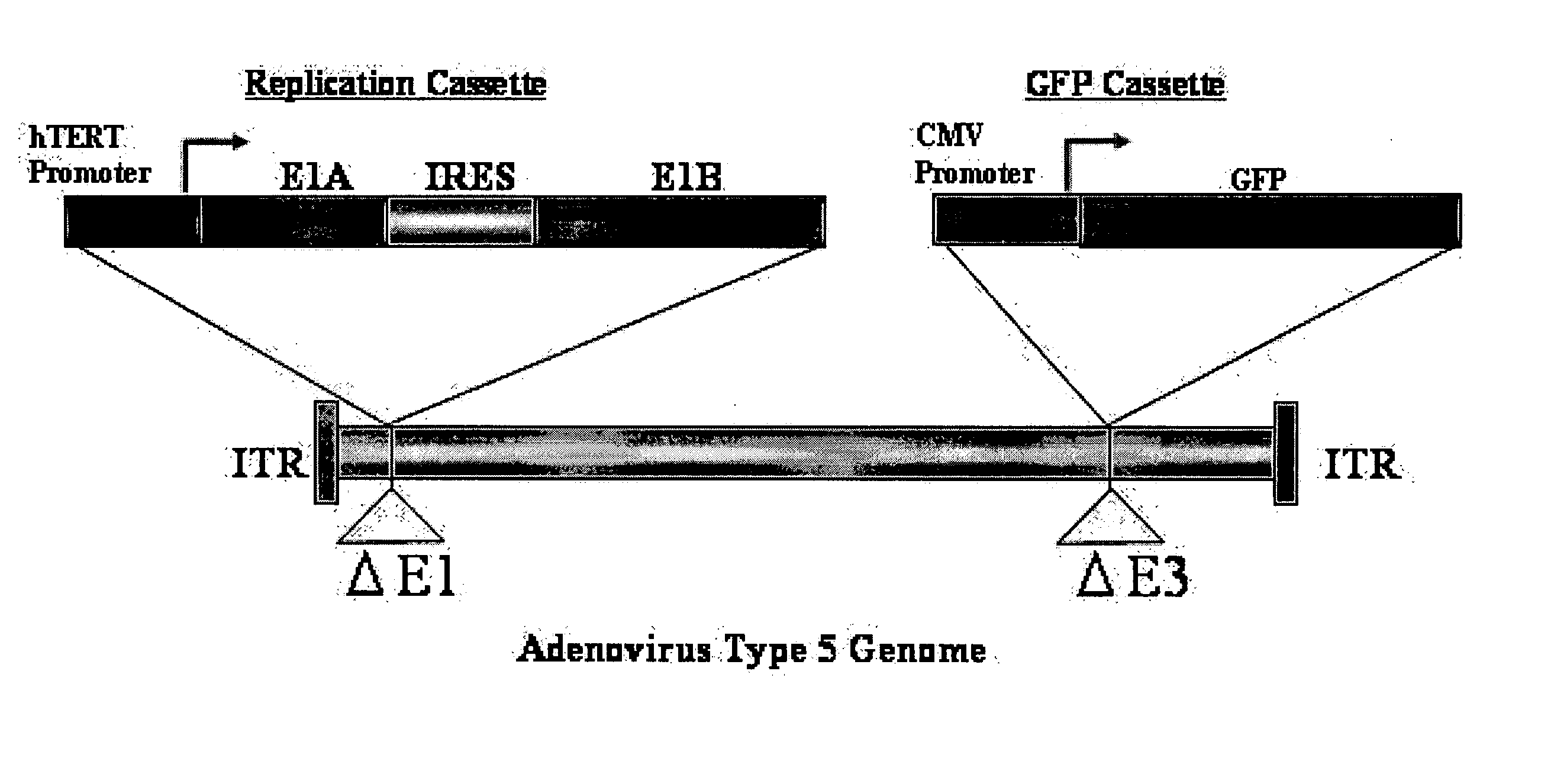
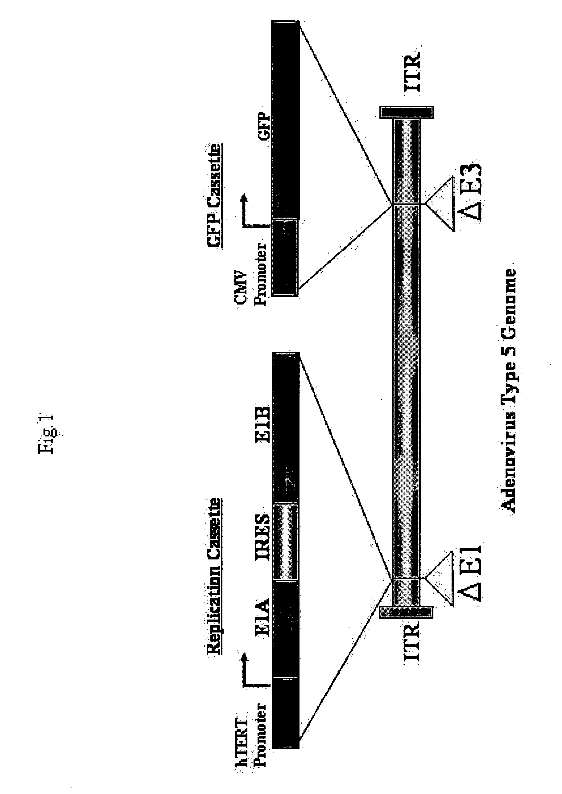
1. Preparation of GFP-Expressing, Replication-Competent Virus (Telomelysin-GFP) Which Comprises the Replication Cassette Comprising Telomerase Promoter and E1 Gene and the Labeling Cassette Comprising Gene Encoding GFP in a Single Virus

[0110] The outline of Telomelysin-GFP is shown in FIG. 1. Telomelysin-GFP proliferates / replicates cancer cell-specifically and telomerase-specifically because E1A / IRES / E1B is operated by the hTERT promoter. Further, Telomelysin-GFP also has an Aequorea victoria-derived GFP gene integrated in its E3 region which is operated by a promoter. Therefore, cells where viral replication is observed emit green fluorescence when excitation light is applied, which enables visualization of cancer cells.

[0111] Such a replication-incompetent virus was prepared as described below.

2. Preparation of Recombinant Virus

[0112] An E1A gene of 897 bp was amplified from RNA extracted from 293 cells by RT-PCR using the follo...


PUM

PropertyMeasurementUnit
Cell deathaaaaaaaaaa
Fluorescenceaaaaaaaaaa

Abstract

The present invention provides a reagent for cancer cell detection or cancer diagnosis. The present invention relates to a reagent for cancer cell detection, comprising a recombinant virus where a replication cassette comprising a promoter from human telomerase, an E1A gene, an IRES sequence and an E1B gene in this order is integrated in E1 region of the viral genome and a labeling cassette comprising a gene encoding a labeling protein and a promoter capable of regulating the expression of the gene encoding the labeling protein is integrated in E3 region of the viral genome.

Description

BACKGROUND OF THE INVENTION [0001] The present invention relates to a reagent for detecting cancer cells or diagnosing cancers, and a cell death-inducing agent. [0002] Telomerase activity is often enhanced in malignantly transformed cells or immortalized cell strains, whereas telomerase activity is hardly detected in normal somatic cells excluding such as germ line cells, blood lineage cells and epithelial cells. Therefore, attempts to detect cancer using telomerase activity as an indicator have been made (Shay J W, Zou Y, Hiyama E, Wright W E. Telomerase and Cancer. Hum Mol Genet 10 (7): 677-85, 2001). [0003] On the other hand, detection of cancer tissues and metastatic lymph nodes in vivo has been studied eagerly in the field of diagnostic imaging. For example, biological diagnosis with PET and image analysis fully utilizing neural network have been reported. Further, investigations into the anti-tumor activity and safety of replication-selective viruses have been reported (DeWees...

Claims


Application Information

Patent Timeline
IPC IPC(8): A61K49/00A61K48/00C12N15/861A61K35/76A61K35/761A61K35/768A61K38/51A61P35/00A61P35/02A61P35/04A61P43/00C12N7/00C12N7/01C12N15/09C12N15/86C12Q1/04G01N21/78G01N33/48G01N33/58
CPCA61K49/0047C07K14/4702C07K14/70575C12N7/00G01N33/574C12N2710/10343C12N2830/008C12N2840/203C12N15/86A61P35/00A61P35/02A61P35/04A61P37/00A61P43/00C12Q1/04A61K48/00C12N15/861C12N15/09
Inventor FUJIWARA, TOSHIYOSHITANAKA, NORIAKIKYO, SATORUMIZUGUCHI, HIROYUKIHAYAKAWA, TAKAO
Owner ONCOLYS BIOPHARMA
Why Eureka
Try Eureka