Method for producing decalcified hard tissue sample

a hard tissue and sample technology, applied in the field of decalcification hard tissue samples, can solve the problems of high sample production cost, insufficient scaffolding, and difficulty in observing the true conditions of cells and overall hard tissue structure, and achieve the effect of keeping the fine structure of hard tissue and low cos

Inactive Publication Date: 2006-03-30
ASAHI KOGAKU KOGYO KK
View PDF0 Cites 1 Cited by
  • Summary
  • Abstract
  • Description
  • Claims
  • Application Information

AI Technical Summary

Benefits of technology

[0006] Accordingly, an object of the present invention is to provide a method for producing a decalcified hard tissue sample simply at a low cost while keeping the fine structure of the hard tissue.

Problems solved by technology

Particularly when living body components are contained in small amounts, however, this method has difficulty in observing the true conditions of cells and the overall hard tissue structures, because the hard tissues are too weak or lost after the calcium components are dissolved away.
In an artificial bone, to which cells are attached and cultured, for instance, it does not have sufficient scaffold because of too little collagen, etc. after decalcification, failing to sufficiently keep the conditions of the attached cells.
This method, however, can produce only as thick sections as about 100 μm, needing the grinding of the sections, and thus resulting in a high sample production cost.
In addition, it suffers the problem that the number of samples obtained from a specimen of the same size is 1 / 100 or less that of decalcified samples.
However, because an embedded hard tissue is sliced in the method of JP 2000-346770 A, the fine structure of the hard tissue would likely be broken by slicing if the hard tissue had high hardness.
In addition, embedding takes 3-4 weeks in this method.
Though the method of JP 2002-31586 A works well on a soft tissue sample, it destroys the fine structure of a hard tissue during slicing.
In addition, it needs a freeze-slicing apparatus, resulting in a high cost.

Method used


Image

  • Method for producing decalcified hard tissue sample
  • Method for producing decalcified hard tissue sample
  • Method for producing decalcified hard tissue sample

Examples


example 1

[0055] Primary osteoblasts obtained from a skull bone of a newborn rat were attached to hydroxyapatite having a diameter of 5 mm, a thickness of 2 mm, and a porosity of 50% before attaching the cells, and cultured. The cell-attached hydroxyapatite was immersed in a 4-%-by-mass formaldehyde / phosphoric acid buffer solution kept at room temperature for 1 week, to fix the cell tissue to the hydroxyapatite. The fixed specimen was washed with flowing water, immersed in aqueous ethanol solutions of 70% and 96%, respectively, by volume at room temperature for 2 hours each, and then immersed in anhydrous ethanol at room temperature for 1 hour for dehydration.

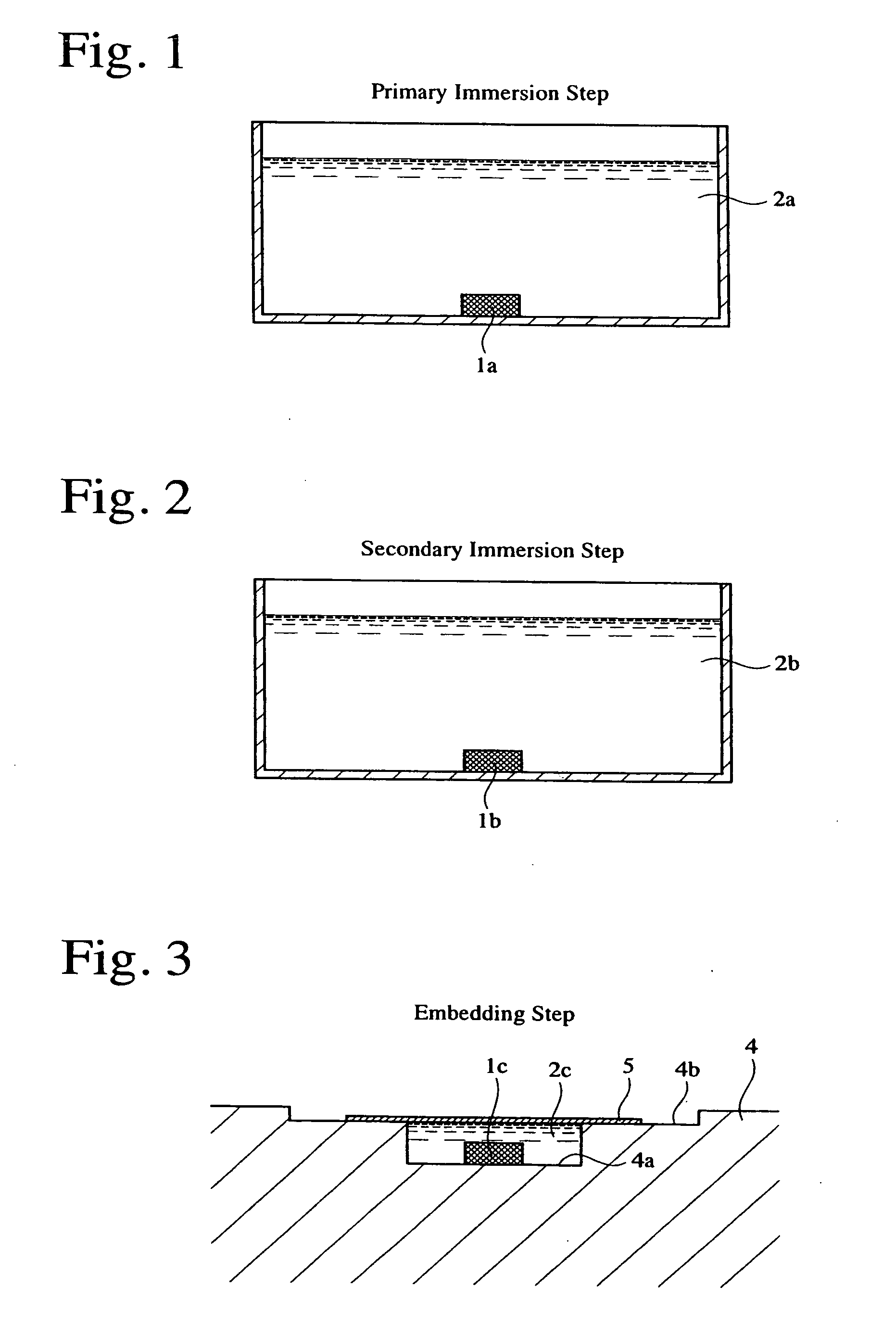
[0056] The dehydrated specimen was immersed in a primary immersion liquid 2a comprising a main ingredient of Technovit 7100 mainly composed of HEMA and including an auxiliary catalyst capable of generating chloride ions, and anhydrous ethanol at an equal volume ratio, at room temperature for 2 hours. The primarily immersed hard tissue p...

example 2

[0060] A sliced sample of cell-containing, decalcified hydroxyapatite was produced in the same manner as in Example 1, except for using ultra-porous hydroxyapatite (HAp-S) having a diameter of 5 mm, a thickness of 2 mm, and a porosity of 85% before attaching the cells, to which commercially available clonal osteoblasts [HOS (human osteosarcoma) cells] were attached and cultured. FIGS. 15-17 are optical photomicrographs of the resultant sample.

example 3

[0061] A sliced sample of cell-containing, decalcified hydroxyapatite was produced in the same manner as in Example 2 except for dying it with toluidine blue. FIGS. 18 and 19 are optical photomicrographs of the resultant sample.

[0062] As shown in FIGS. 15-19, even when the ultra-porous hydroxyapatite was used, a fine tissue structure was clearly observed as in the case of using hydroxyapatite having usual porosity (Example 1).


PUM


Abstract

A method for producing a decalcified hard tissue sample by embedding a hard tissue in a liquid-penetration-permitting resin, and then decalcifying it.

Description

FIELD OF THE INVENTION [0001] The present invention relates to a method for producing a decalcified hard tissue sample, particularly to a method for producing a decalcified sample of a living hard tissue replacement, a living hard tissue or their composite, to which cells are attached, simply at a low cost. BACKGROUND OF THE INVENTION [0002] To microscopically observe hard tissues such as composites comprising scaffolds constituted by living hard tissue replacements, to which cells are attached, living hard tissues, etc., samples of 10 μm or less in thickness are usually produced. Such samples have conventionally been produced by decalcifying hard tissues, embedding them in paraffin, slicing them, and staining them. Particularly when living body components are contained in small amounts, however, this method has difficulty in observing the true conditions of cells and the overall hard tissue structures, because the hard tissues are too weak or lost after the calcium components are d...

Claims


Application Information

Patent Timeline
Patent Type & Authority Applications(United States)
IPC IPC(8): A61F2/00
CPCA61L27/12A61L27/3687A61L2430/40A61L27/425A61L27/56A61L27/3804
Inventor OKIHANA, HIROYUKIMUKAIDA, MASAHIRO
Owner ASAHI KOGAKU KOGYO KK
Why Eureka
Try Eureka