
Method for screening cells and method for detecting oral carcinoma cells

a cancer cell and cell technology, applied in the field of oral carcinoma cell screening, can solve the problems of not finding a method for early diagnosis of oral carcinoma, and it is almost impossible to evaluate the degree of cancer malignancy before starting treatment, and achieve the effect of high accuracy and effective method for evaluating the degree of cancer malignancy

Inactive Publication Date: 2006-03-30
CANON KK
View PDF21 Cites 5 Cited by
  • Summary
  • Abstract
  • Description
  • Claims
  • Application Information

AI Technical Summary

Benefits of technology

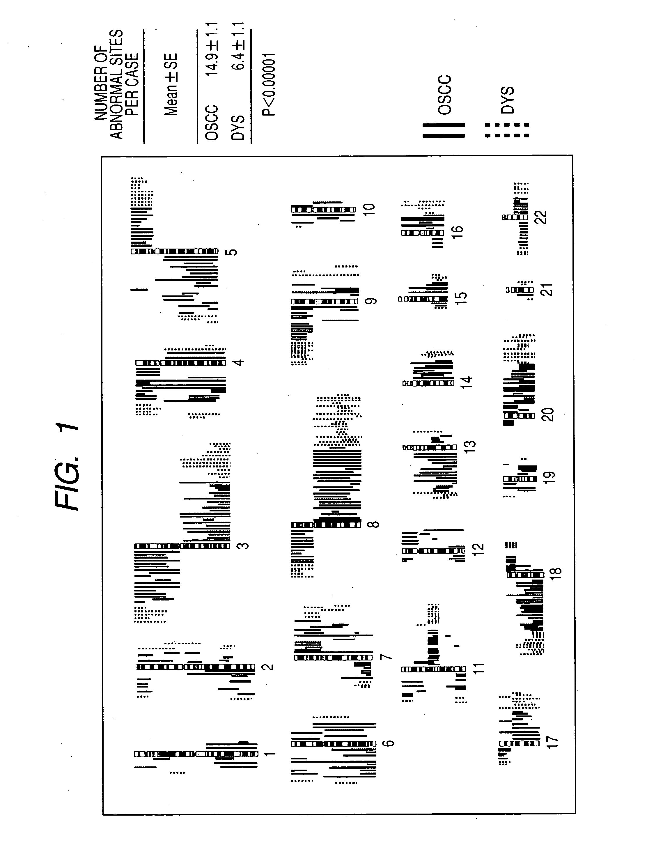
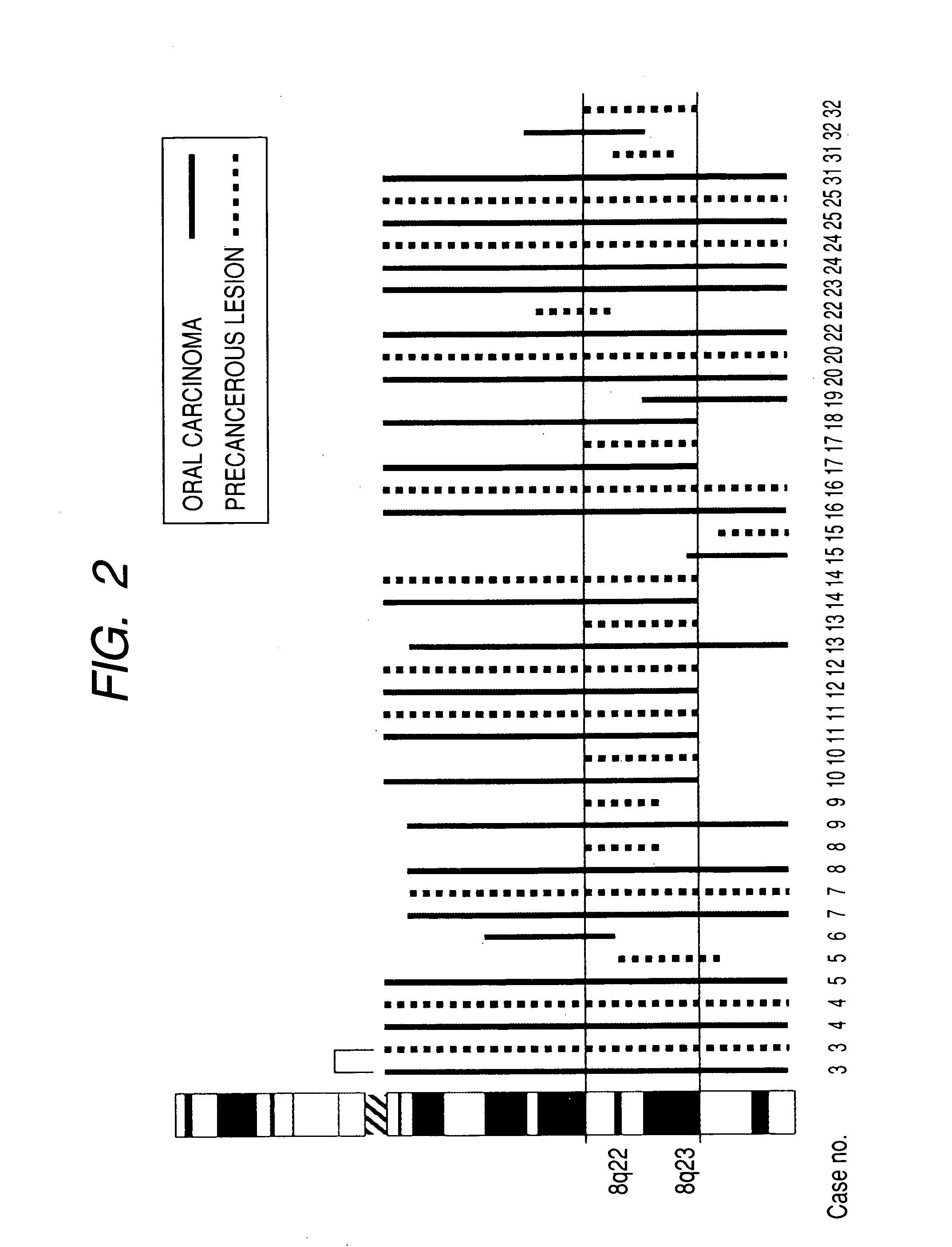
The present invention is about a method for early diagnosis and evaluation of malignancy of oral carcinoma using chromosomal DNA analysis. The inventors have discovered specific chromosomal regions where abnormal DNA copy number occurs in oral carcinoma cells, which can be used for early detection and discrimination between precancerous lesion and early cancer. The method measures the copy number of chromosomal DNA and compares it with normal healthy people. The invention also provides a DNA array for detecting oral carcinoma cells and an effective method for evaluating the degree of malignancy of cancer. Overall, the invention provides a reliable and accurate tool for diagnosis and treatment of oral carcinoma.

Problems solved by technology

As described above, attempts have been made to analyze abnormality in the DNA copy number for cancer diagnosis, but no method for early diagnosis for oral carcinoma is found yet, because the chromosomal region where abnormality in the DNA copy number occurs in oral carcinoma cells is not yet found.
Also, at this time it is almost impossible to evaluate the degree of malignancy of the cancer before starting the treatment.

Method used


Image

  • Method for screening cells and method for detecting oral carcinoma cells
  • Method for screening cells and method for detecting oral carcinoma cells

Examples


example 1

Screening of Cells by the CGH Method

1. DNA Extraction

[0056] To maximize the detection sensitivity of abnormality in the CGH method, samples were prepared by the microdissection method to collect cancer cells selectively.

[0057] In particular, biopsy samples were stored at −80° C., and tissue sections with 9 μm thickness were prepared. After staining with methyl green or hematoxylin-eosin, the tissue section was recovered as 15 μm spots by laser capture dissection and extracted with SepaGene Kit (Sanko Junyaku Co.) to obtain DNA.

2. DOP-PCR

[0058] DOP (degenerate oligonucleotide-primed)-PCR was carried out using a universal primer-6-MW (5′-CCGACTCGAGNNNNNNATGTGG-3′).

[0059] In particular, to 1 μl of DNA obtained by microdissection, 4 μl of sequenase buffer and 1 U of topoisomerase (Promega Co. Trade Mark, Madison, Wis.) were added and incubated at 37° C. for 30 min. Next, the reaction mixture was mixed with 20 U of Thermosequenase (Amersham Co. Trade Mark, Cleaveland, OH) and su...

example 2

Preparation of BAC Clone Microarrays

[0076] Slide glasses were washed and treated with aminosilane coupling agent according to the conventional method.

[0077] The clones corresponding to the chromosomal regions, which were determined to be useful for screening of oral carcinoma based on the result of the analysis in Example 1 (Table 1), were selected from the BAC clone library prepared by the conventional method. These clone solutions were injected into a 96 well microtiter plate and spotted on the slide grass with a DNA microarray apparatus. After standing at room temperature and fixing, the microarray was subjected to competitive hybridization reaction with DNAs of normal healthy people and samples labeled with different fluorescent dyes to obtain the ratio of copy numbers of DNAs of samples and normal healthy people. By defining the ratio of 1.2 or above as amplification and 0.8 or below as loss, the results obtained were almost the same as Example 1.

example 3

Preparation of the Back Clone Microarray with DNA of Normal Healthy People Fixed Beforehand

[0078] A back clone microarray was prepared as described in Example 2, and DNA of lymphocytes of normal healthy people, which was labeled with Spectrum Red, was fixed thereto by hybridization.

[0079] Sample DNA was labeled with Spectrum Green and hybridized with the microarray in which DNA of normal healthy people was already bound to back clones.

[0080] The red fluorescence of DNA of normal healthy people, which was originally bound to back clones, was replaced with the green fluorescence derived from the sample, and the ratios between the two were similar to those in Example 2.


PUM

PropertyMeasurementUnit
thicknessaaaaaaaaaa
temperatureaaaaaaaaaa
frequencyaaaaaaaaaa

Abstract

The present invention provides a method of detecting oral carcinoma cells with high accuracy, and a method of making possible early detection of oral carcinoma or discrimination between a precancerous lesion and an early cancer in diagnosis of oral carcinoma. The methods are achieved by screening cells for oral carcinoma or precancerous lesion by measuring a DNA copy number in whole chromosomes or a part thereof in a sample, wherein chromosomal regions for which said copy number is measured comprise at least one region selected from the group consisting of: a 22-23 region in the q arm of Chromosome 8, a 14-21 region in the p arm of Chromosome 3 and a 12-22 region in the p arm of Chromosome 5.

Description

BACKGROUND OF THE INVENTION [0001] 1. Field of the Invention [0002] The present invention relates- to a method for screening oral carcinoma cells. The present invention also relates to a method for discriminating between cells in precancerous condition (precancerous lesion cells) and oral carcinoma cells, and detecting oral carcinoma cells early. [0003] 2. Related Background Art [0004] Oral carcinoma is estimated to be the 6th most frequent cancer in the world and occurs at high frequency especially in certain areas of Asia. Oral carcinoma includes cancer of the maxillary sinus, which is located inside the left and right cheek, cancer of the tongue in the mouth, cancer of the epipharinx in the nose or in the deep throat, cancer of the larynx in the periphery of the vocal cord and the like. In spite of the fact that they are very common cancer, the prognosis is not good, and there are frequent recurrences and in many cases, death is the result. One of the reasons for these is the dif...

Claims


Application Information

Patent Timeline
Patent Type & Authority Applications(United States)
IPC IPC(8): C12Q1/68
CPCC12Q2600/112C12Q1/6886
Inventor YAMAMOTO, NOBUKOSASAKI, KOHSUKE
Owner CANON KK
Learn More