Apparatus and methods for steroid hormone testing

a technology of steroid hormone and steroid hormone, applied in the field of steroid hormone testing, can solve the problems of bone loss, slow process, osteoporosis, etc., and achieve the effect of preventing bone loss, preventing bone loss, and preventing bone loss

Inactive Publication Date: 2006-03-30
NJ INT
View PDF6 Cites 16 Cited by
  • Summary
  • Abstract
  • Description
  • Claims
  • Application Information

AI Technical Summary

Problems solved by technology

But once estrogen levels start to decline, this process also slows down.
Although bone loss eventually levels out more than five years post-menopause, keeping bone structures strong and healthy to prevent osteoporosis becomes more of a challenge.
Osteoporosis occurs when bones become too weak and brittle to support normal activities.
The long-term health consequences of estrogen decline after menopause includes bone loss, increased risk of cardiovascular disease, and cognitive impairment.
Recently, however, it was reported that women who started ERT at a later age are under an increased risk for developing breast cancer.
But due to aging, body fat, hormonal replacement, pesticides, nutritional deficiencies, prescription medications and excessive alcohol intake many men experience high levels of estrogen which are detrimental to their health.
A testosterone / estrogen imbalance directly causes many of the debilitating health problems associated with normal aging.
Therefore, accurate detection of steroids in biological samples generally require highly sensitive methods.
Detecting estrogens in a biological fluid from postmenopausal women, children or men is particularly demanding on the method's sensitivity where concentrations at the low pg / ml level are encountered.
U.S. Pat. No. 5,342,760 discloses and claims a useful method for determination of estradiol in fluid samples by competitive immunoassay, but these methods typically require professional training or specialized equipment, and take a long time to complete.

Method used


Image

  • Apparatus and methods for steroid hormone testing
  • Apparatus and methods for steroid hormone testing

Examples


example 1

Preparation of Binding Membrane

1. Preparation of Colloid Gold Solution

[0056] Prepare the glass vessels by soaking in 3%-10% dimethyldichlorosilane chloroform solutions for about 1 minute, air dry, washing with the distilled water, and air dry again at room temperature. Mix 80-120 ml of 0.08%-0.12% chloroauric acid solution with 0.5-0.9 ml of 0.8%-0.12% sodium citrate in a preheated glassware, heat to boiling. The solution will turn from yellow to purple. Continue to boil for 10-20 minutes. After cooling, add distilled water to bring the volume to the original (80-120 milliliter).

2. Preparation of Colloid-Gold Labeled Antigen (Estradiol)

[0057] The protocol is as follows: 1. Adjust colloid gold solution prepared in Example 1 to pH 8.2-8.6 using 0.08-0.12 M potassium acetate solution; 2. Mix 300-500 μg of antigen (estradiol) with 80-120 ml colloid gold solution for 10-15 min at room temperature; 3. Add 4-10 ml of 0.8-1.3% polyethylene glycol solution; 4. Centrifuge at 10,000˜100,...

example 2

Preparation of Nitrocellulose Binding Membrane

[0060] On a strip of nitrocellulose membrane, apply an antibody against the analyte antigen (estradiol) in three detection lines. The detection lines are about 1-3 mm wide, and are about 2-3 mm apart. From the end that is closest to the sample membrane, the coating concentrations of antibody on the nitrocellulose membrane are 0.4, 1.2, 3.0 and, 4.8 μg, respectively, which correspond to an estrogen concentration of 50, 200, 500, 1200 pg / ml in a 100 μl sample. The standard curve is established using the rate of 4 methylumbelliferone formation. A total of 4 testing lines and a 5 μg control antibody are used. A fifth line, of the anti-IgG antibody, which is also about 2-3 cm from the detecting line next to it, is also applied on the nitrocellulose membrane.

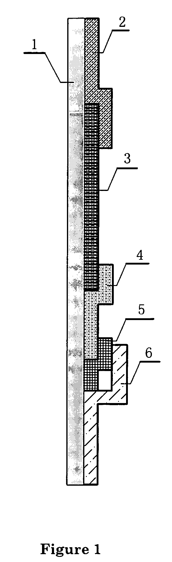
[0061] The final chromatographic test device or “strip” is prepared as shown in FIG. 1. It includes 1 as supporting plate, 2 as absorbing pad, 3 as the nitrocellulose binding membrane, 4...

example 3

Detection and Quantification of Estrogen Level in a Sample Using the Chromatographic Analysis Device

[0062] A sample of about 0.1-0.5 ml saliva or urine is applied (for example, using a tube provided with the kit, 1 drop is about 50 μl), without any pre-treatment or preparation, to the sample membrane, and allowed to react for 1 min. Then the water-proof membrane between the sample membrane and the colloid gold membrane is removed, following by addition of 0.1 ml of water on the colloid gold membrane. Allow two minutes for the labeled antigens to migrate through the binding membrane. Afterwards, the nitrocellulose membrane is examined visually to determine if any of the detection line has undergone any color change (i.e. turning red). If all four detection lines turn red, then the sample contains less than 50 pg / ml of estrogen. If three four detection lines turn red, then the sample contains about 50 pg / ml of estrogen. If two detection lines turn red, then the sample contains about ...


PUM

PropertyMeasurementUnit
thickaaaaaaaaaa
thickaaaaaaaaaa
molecular weightaaaaaaaaaa

Abstract

An immuno-chromatographic detection device for detecting an analyte in sample, such as estrogen in a urine or saliva sample, the device comprising (a) a binding membrane having immobilized thereon (i) an test antibody against said analyte in at least one detection region, and (ii) a control antibody against a control antigen known to be present in the sample in a control region, (b) a sample membrane located at a first end of the binding membrane for receiving the sample, wherein the sample membrane is in chromatographic connection with the binding membrane, and (c) a label membrane containing (iii) a labeled antigen that is capable of binding to the test antibody and upon binding with the test antibody exhibits an observable change at the at least one detection region, and (iv) a labeled control antigen that is capable of binding to the control antibody and upon binding with the control antibody exhibits an observable change at the control region, wherein the sample membrane is separated from the label membrane by a waterproof membrane which is removable to allow the sample membrane and label membrane to be connected chromatographically. Also provided are kits comprising the device, method for detecting the analyte, and methods for manufacturing the device and kit.

Description

FIELD OF THE INVENTION [0001] This invention relates to apparatus and methods for determining the concentration of a steroid hormone in a human subject. BACKGROUND OF THE INVENTION [0002] Estrogens are small-molecule steroids synthesized in a number of human cells and tissues, such as ovarian granulose cells, placental syncytiotrophoblast, adipose tissue and the brain. Estrogens include typically estradiol, estriol, and estrone. Estradiol, the principal hormone of the ovary, is important for female sexual differentiation during gestation, sexual development at the onset of puberty, and regulation of the menstrual cycle. Estradiol plays roles in the menstrual cycle and involved in fertilization by both the stimulation and inhibition of the release of the gonadotropins, exerting both a positive and a negative feedback. Early in the follicular phase, ovarian secretion of estradiol from the thecal and granulosa cells is modest. During the follicular phase, estradiol stimulates endometri...

Claims


Application Information

Patent Timeline
Patent Type & Authority Applications(United States)
IPC IPC(8): G01N33/558
CPCG01N33/743G01N33/558G01N33/54388
Inventor LI, RENAYANG, LIBANGSHEN, YONG
Owner NJ INT
Why Eureka
Try Eureka