Ultrasound-activated anti-infective coatings and devices made thereof

a technology of anti-infection coating and ultrasonic activation, which is applied in the direction of catheters, food packaging, therapy, etc., can solve the problems of port removal, increased infection risk, and use of cvads

Active Publication Date: 2006-05-11
BOSTON SCI SCIMED INC
View PDF31 Cites 39 Cited by
  • Summary
  • Abstract
  • Description
  • Claims
  • Application Information

AI Technical Summary

Problems solved by technology

However, an added risk of infection exists due to the presence of the exterior component.
Risks associated with the use of CVADs include local complications such as thrombosis and thrombophlebitis, as well as systemic complications including embolisms, pulmonary edema and bloodstream infections.
During these two years, infection will develop in around 20% of patients, and often leads to removal of the port.
Infection of the A-V port has been recorded as a major cause of death in patients receiving dialysis treatments.
Second, bacteria may already be present on the surface of the device.
It is this virulence that leads to septicemia and is one main factor in the high mortality rates.
Vascular port infections are difficult to treat with the standard course of oral antibiotics.
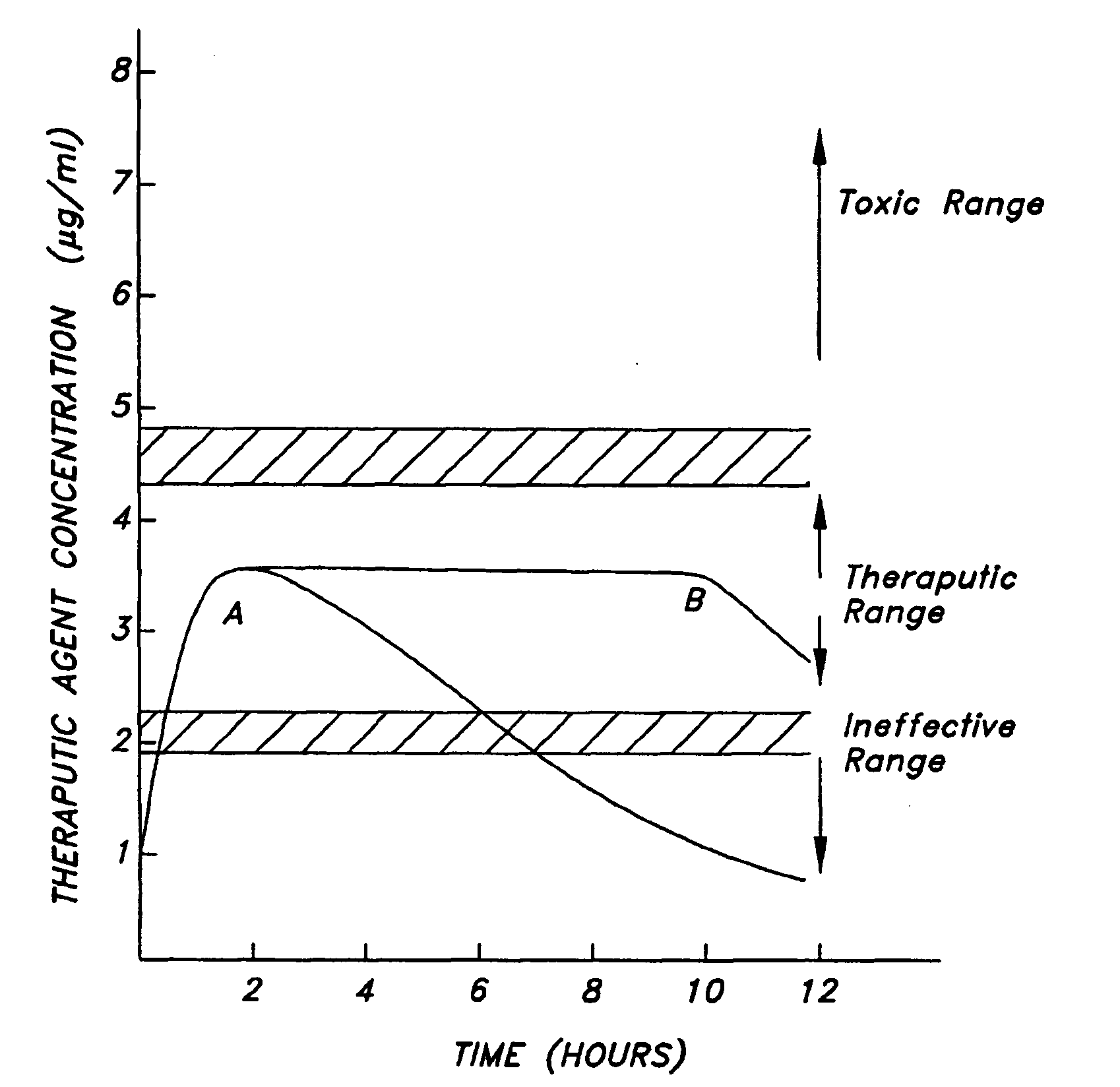
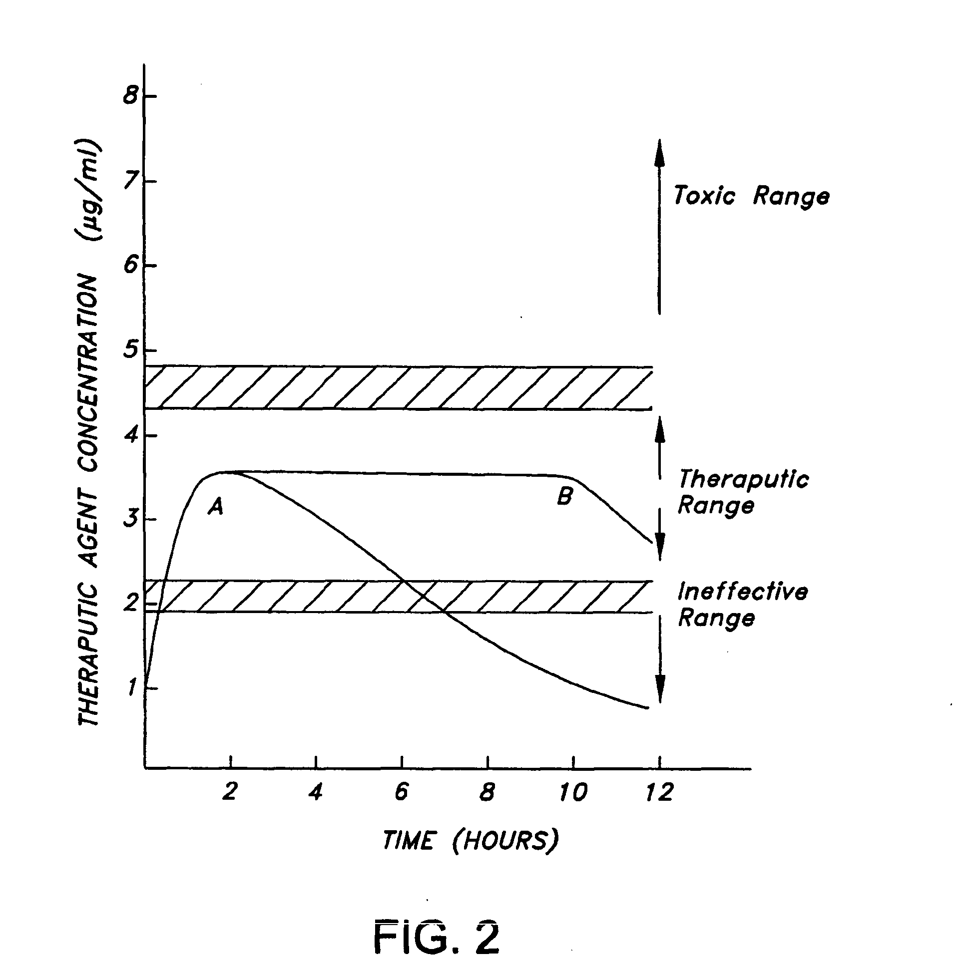
A disadvantage of presently available methods for providing therapeutic agents on medical device substrates is the lack of a means to control the rate of release of the therapeutic agent.
Furthermore, an active infection may require a larger dose than is delivered by sustained release of the therapeutic (i.e. anti-microbial) agent.

Method used


Image

  • Ultrasound-activated anti-infective coatings and devices made thereof
  • Ultrasound-activated anti-infective coatings and devices made thereof
  • Ultrasound-activated anti-infective coatings and devices made thereof

Examples


example 1

Ultrasound Activatable Micelles

[0124] This example describes a coating made with a cross-linked polymeric material formed into micelles loaded with therapeutic agent that will release the agent upon exposure to ultrasound energy. An amphiphylic alternating copolymer consisting of poly(ethyleneglycol) and poly(L-lactic acid) as shown below is used to form micelles. The polymeric micelles are further stabilized by polymerization using N,N-diethylacrylamide in a poly(L-lactic acid) inner core of the micelles. The micelles are further optimized by reaction of acetylated hydroxyalkyl carboxylic acid derivatives to add functional groups, such as —COOH, SO4, H, NH or the like, as attachment sites for the therapeutic agent.

[0125] The hydrophilic / hydrophobic copolymer is dissolved in methylene chloride and emulsified with a 5% aqueous solution of albumin containing an antibiotic and / or a thrombogenic agent by sonicating for 2 minutes, and spray-drying to produce particles (10 μm). The mic...

example 2

Ultrasound Activatable Micelles

[0126] This example describes a coating made with a cross-linked polymeric material which forms micelles loaded with therapeutic agent that will release the agent upon exposure to ultrasound energy. Poly(ethylene oxide glycol) / poly(propylene oxide glycol) copolymers and poly(Ε-capriolactone) are used to form a block polymer as shown below.

The core of this polymeric micelle is stabilized by forming an interpenetrating cross-linked system using N,N,diethylacrylamide as a cross-linking agent. The therapeutic agent is incorporated into the micelle as described above. The micelle is dried and used directly as a coating on a medical device substrate or is dried and applied onto a polymeric coating on the substrate.

example 3

Ultrasound Activatable Micelles

[0127] This example describes a coating made with a cross-linked polymeric material which forms micelles loaded with therapeutic agent that will release the agent upon exposure to ultrasound energy. A commercially available poly(ethylene / glycol)-poly(propylene glycol) triblock copolymer (PEO-PPO-PEO), as shown below is used to make the polymeric material of the coating. The copolymer is optionally cross-linked to form an interpenetrating network.

To a commercial polymer, (PLURONIC 105 or PLURONIC 127, available from BASF Corp., Ludwigshafen, Germany) is added N,N-diethylacrylamide to stabilize the hydrophobic core of the micelle. The therapeutic agent is incorporated into the micelle as described above. The micelles are dried and used directly as a coating on a medical device substrate or are dried and appied onto a polymeric coating on the substrate.


PUM

PropertyMeasurementUnit
wavelength rangeaaaaaaaaaa
timeaaaaaaaaaa
pore sizeaaaaaaaaaa

Abstract

An implantable medical device is provided including a vascular access device and a coating on at least one of an inner surface and an outer surface of the vascular access device. The coating includes: (a) a polymeric component including at least one of a light reactive moiety and a sound reactive moiety; and (b) at least one therapeutic agent releasably associated with the polymeric component, wherein a rate of release of the therapeutic agent is controlled by in situ exposure of the medical device to at least one of a light energy source and an ultrasound energy source.

Description

FIELD OF THE INVENTION [0001] The present invention relates generally to implantable medical devices having a biocompatible polymer coating for delivery of therapeutic agents. More particularly, the present invention relates to an implantable medical device having a biocompatible polymer coating including at least one therapeutic agent whereby the therapeutic agent is released from the coating by exposure to at least one of ultrasound energy and light energy. BACKGROUND OF THE INVENTION [0002] Central vascular access devices (CVADs) are medical devices that are implanted into a patient's vascular system and are typically used in applications which provide a means for repeated access to a patient's vascular system. Applications for CVADs are varied and include, for example, intravenous feeding, intravenous drug delivery, and extracorporeal protocols. Specific applications include chemotherapy treatments, intensive antibiotic treatment, prolonged IV feeding, and extracorporeal blood t...

Claims


Application Information

Patent Timeline
Patent Type & Authority Applications(United States)
IPC IPC(8): A61N1/30A61M31/00
CPCA61K41/0033A61K41/0042A61L29/085A61L29/16A61L31/10A61L31/16A61L2300/404A61L2300/416A61L2300/602
Inventor TAN, SHARON MI LYN
Owner BOSTON SCI SCIMED INC
Why Eureka
Try Eureka