Method and system for registering an image with a navigation reference catheter

a technology of image data and navigation reference, which is applied in the field of medical probes, can solve the problems of inability of physicians to visualize anatomy features as a reference for navigation, lack of resolution of conventional imaging systems, and inability of physicians to use fluoroscopy to locate catheters, etc., to achieve the effect of reducing errors and time, registering and merging graphical information and image data can be more accura

Active Publication Date: 2006-06-01
BOSTON SCI SCIMED INC
View PDF14 Cites 207 Cited by
  • Summary
  • Abstract
  • Description
  • Claims
  • Application Information

AI Technical Summary

Benefits of technology

[0020] Registration and merger of the graphical information and image data can be made more accurate by correlating another physical structure with the functional catheter. For example, the method may comprise locating the other physical structure spatially correlated to the functional catheter, identifying another image reference corresponding to the other physical structure, and merging the graphical information and medical image further based on the other located physical structure and the other identified image reference.
[0021] In accordance with a fourth aspect of the present inventions, a medical system for superimposing the medical image and graphical information is provided. The system comprises a reference catheter, a functional catheter, and control circuitry (e.g., ranging circuitry) configured for conditioning the reference and functional catheters to transmit signals (e.g., ultrasound pulses) between each other. The system further comprises one or more processors configured for acquiring location information with the functional catheter based on the transmitted signals, locating a physical structure spatially correlated to the reference catheter, generating the graphical information based on the location information, identifying an image reference corresponding to the physical structure, and merging the graphical information and medical image based on the located physical structure and the identified image reference. These functions can be performed in the same manner described above. Registration and merger of the graphical information and medical image can be made more accurate as described by correlating another physical structure with the functional catheter in the manner described above.
[0022] In accordance with additional aspects of the present inventions, methods and medical systems for processing the medical image and / or superimposing the medical image and graphical information, are provided. These methods and systems are similar to those methods and systems described above, with the exception that the reference element(s) and / or reference catheter need not be used to establish a navigational coordinate system or otherwise used to communicate with location elements or catheters. However, the methods and systems in accordance with these additional aspects of the present inventions do require that image reference be identified by automatically analyzing the image data to identify reference data corresponding to the physical structure, which is then used to register the coordinate systems. Thus, any errors and time expended as a result of a manual process of identifying a reference with the medical image is eliminated or at least minimized.

Problems solved by technology

The use of fluoroscopy in locating catheters is somewhat limited, however, in that the physician is only able to visualize the catheter and surrounding tissues in two dimensions.
In addition, fluoroscopy does not image soft tissues, making it difficult for the physician to visualize features of the anatomy as a reference for the navigation.
Although the uses of the RPM tracking system and CARTO EP system have, to a large extent, been successful in facilitating the navigation of catheters within the cavities of the heart, the graphical representations of the heart chambers lack the resolution produced by conventional imaging systems, such as Computed Tomography (CT) and Magnetic Resonance Imaging (MRI) systems.
This lack of resolution especially impacts the navigation of catheters within and around the complex anatomy of the left atrium and pulmonary veins.
Besides requiring an additional step to be performed by the user, the image integration is only as accurate as that of the anatomical envelope of the electrophysiological map, which as discussed above, lacks the resolution typically seen in a CT image.
As such, catheter localization relative to the CT image may be inaccurate to a certain extent.
Thus, the CT image cannot be used when initially navigating the catheter within the heart.

Method used


Image

  • Method and system for registering an image with a navigation reference catheter
  • Method and system for registering an image with a navigation reference catheter
  • Method and system for registering an image with a navigation reference catheter

Examples


Embodiment Construction

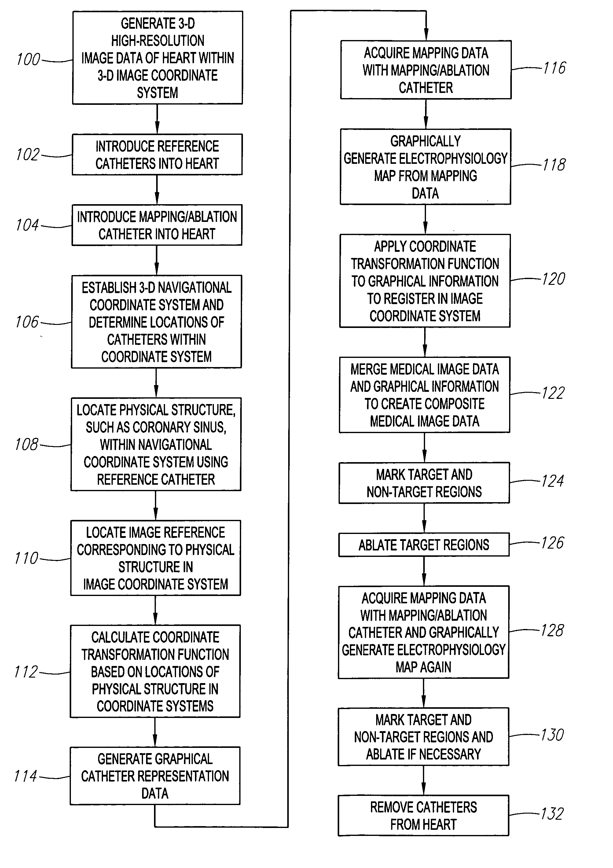
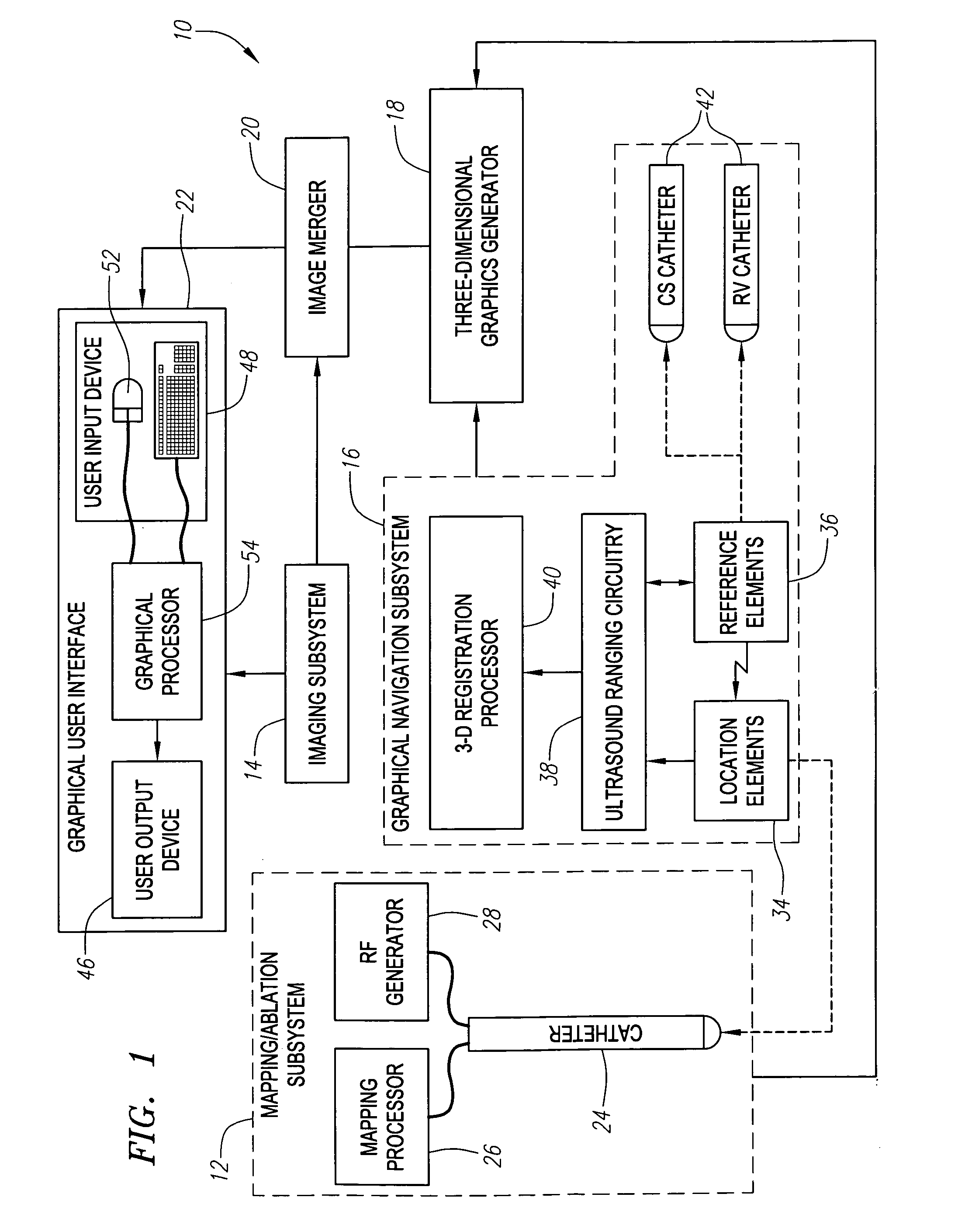
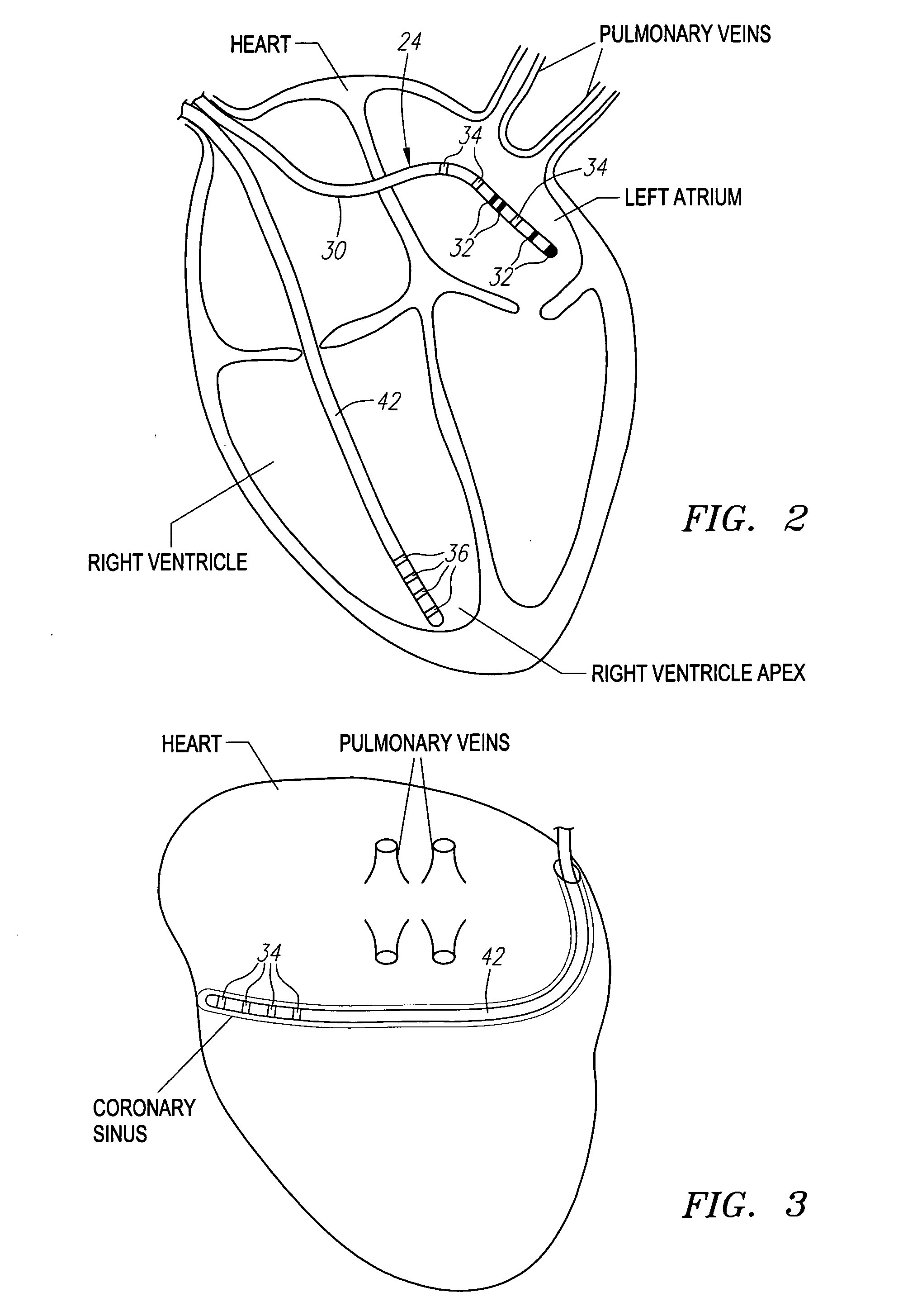
[0033] Referring to FIG. 1, an exemplary medical system 10 constructed in accordance with the present invention is shown. The medical system 10 is particularly suited for mapping and treating a heart (shown in FIGS. 2 and 3). Nevertheless, it should be appreciated that it can be used for treating, diagnosing, or otherwise displaying other anatomical bodies, such as the prostate, brain, gall bladder, uterus, esophagus, and other regions of the body.

[0034] The medical system 10 generally comprises (1) a mapping / ablation subsystem 12 for mapping and ablating tissue within the heart; (2) an imaging subsystem 14 for generating a high-resolution medical image of the heart; (3) a graphical navigation subsystem 16 for registering mapping data and the movement of medical devices within a three-dimensional navigational coordinate system (which in the illustrated embodiment, may also be referred to as a graphical coordinate system); (4) a three-dimensional graphics generator 18 for generating...


PUM


Abstract

Methods and systems for processing and/or superimposing a medical image of an anatomical body (e.g., a heart) with graphical information are provided. Reference elements and/or reference catheter are placed in contact with the anatomical body. A physical structure within a navigational coordinate system is located using the reference elements and/or reference catheter. An image reference within an image coordinate corresponding to the physical structure is located. Location of the image reference can be accomplished, e.g., by displaying the medical image and electronically marking the displayed image reference, or by automatically locating image data corresponding to the image reference. The navigational and image coordinate systems are then registered based on the location of the physical structure within the navigational coordinate system and the location of the image reference within the image coordinate system, which allows graphical information to be accurately merged with the medical image data.

Description

FIELD OF THE INVENTION [0001] The present inventions generally relate to medical probes, and more particularly to systems and methods for navigating catheters within anatomical organs or other anatomical structures. BACKGROUND OF THE INVENTION [0002] It is often necessary or desirable to determine the location of a medical probe relative to a location of interest within three-dimensional space. In many procedures, such as interventional cardiac electrophysiology therapy, it is important for the physician to know the location of a probe, such as a catheter, (especially, a therapeutic catheter) relative to the patient's internal anatomy. During these procedures, a physician, e.g., steers an electrophysiology mapping catheter through a main vein or artery into the interior region of the heart that is to be treated. The physician then determines the source of the cardiac rhythm disturbance (i.e., the targeted cardiac tissue) by placing mapping elements carried by the catheter into conta...

Claims


Application Information

Patent Timeline
Patent Type & Authority Applications(United States)
IPC IPC(8): A61M25/00
CPCA61B5/0422A61B6/12A61B6/503A61B6/5247A61B8/0833A61B8/0841A61B8/0883A61B8/463A61B8/5238A61B19/52A61B2019/5289A61B2019/5429A61B5/743A61B90/36A61B2090/364A61B2090/3929A61B5/287
Inventor WILLIS, N. PARKER
Owner BOSTON SCI SCIMED INC
Why Eureka
Try Eureka