Nanofibrous biocomposite prosthetic vascular graft

a biocomposite, prosthetic technology, applied in the field of vascular prosthesis, can solve the problems of graft failure, difficult suture into proper place, and difficult handling of graft materials, and achieve the effect of improving the quality of vascular grafts

Active Publication Date: 2006-06-15
BIOSURFACES +2
View PDF6 Cites 26 Cited by
  • Summary
  • Abstract
  • Description
  • Claims
  • Application Information

AI Technical Summary

Benefits of technology

[0032] A first aspect provides a fabricated textile conduit useful as a tangible workpiece for the manufacture of a prosthetic vascular graft construct, said fabricated textile article comprising:
[0033] a nanofibrous biocomposite material comprised of at least one biodurable synthetic substance and at least one extracellular matrix protein and fabricated as an elongated, hollow tube via an electrospinning perfusion process, said fabricated textile conduit having determinable inner wall and outer wall diameter sizes, two open ends, and an internal lumen less than about 6 millimeters in diameter, and presenting a discrete interior wall surface and an exterior wall surface, and being biocompatible for the conveyance of blood through its internal lumen.
[0034] A second aspect of the p...

Problems solved by technology

Although early results were encouraging, the long-term results were still unsatisfactory, with the grafts often failing due to thrombosis and aneurysm.
In general, the woven or knitted types of vascular graft provide a greater strength and a reduced porosity for the substrate, but these types of graft materials are considered to be more difficult to handle and to suture into proper place.
Compaction results in the shrinking of the yams comprising the graft article and generally reduces the overall porosity of the fabric substrate.
Note however, that the degree of protection afforded by irregular corrugation varies over the lengths of the tube and can fall below a required level of protection at specific regions.
Tendon-derived collagen is generally highly cross-linked and difficult to process by the enzyme digestion procedure described in the patent.
While accepted for medical use in small diameter applications, PTFE graft constructions often require surgical replacement within relatively short periods of time compared to the larger diameter fabric vascular grafts described above.
Graft articles made using these materials have proven to be successful for large diameter artery replacement where there is a high blood flow rate; but unfortunately they have a much lower success rate in blood vessels with an inner diameter size less than 6 mm.
In addition, the substrates comprising these conventionally used vascular grafts do not permit unrestricted ingrowth from the surrounding tissue after in-vivo implantation, owing mostly to the existence of ingrowth spaces within the substrate material that are either too narrow or are discontinuous.
Although the reasons for failure are not fully understood, it is largely agreed that compliance mismatch between blood vesse...

Method used


Image

  • Nanofibrous biocomposite prosthetic vascular graft
  • Nanofibrous biocomposite prosthetic vascular graft
  • Nanofibrous biocomposite prosthetic vascular graft

Examples


Embodiment Construction

I. The Subject Matter of the Present Invention as a Whole

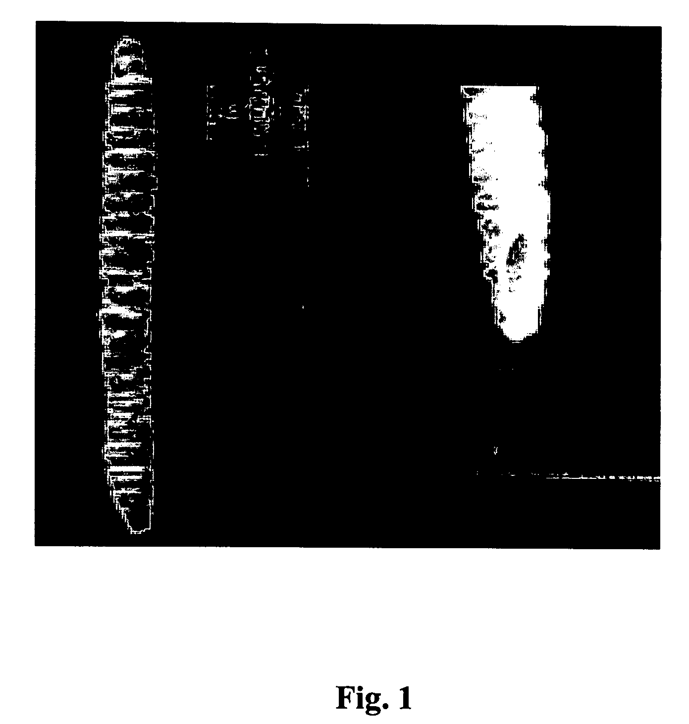
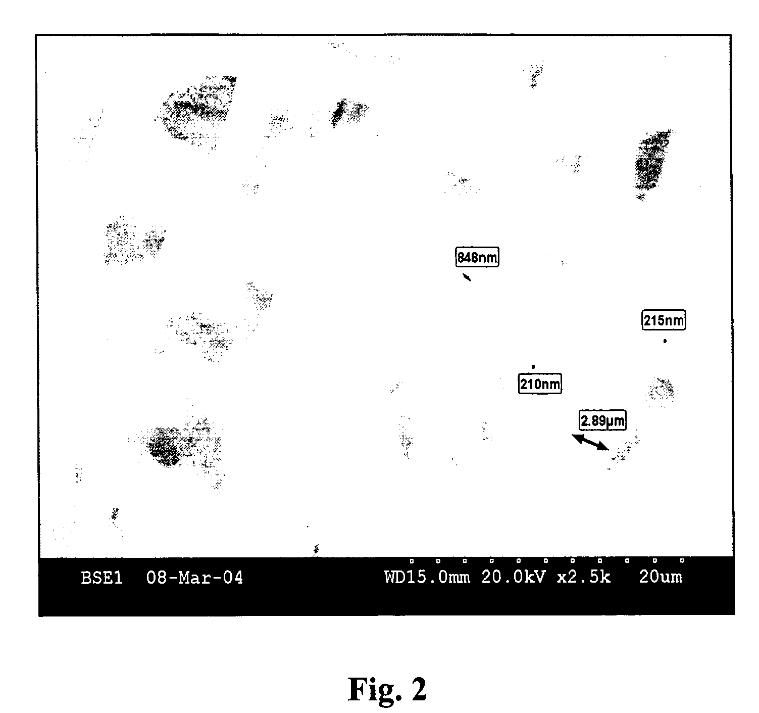
[0085] The present invention comprises a bioactive, small-diameter (typically less than 6 mm in internal diameter) nanofibrous vascular graft prosthesis which is preferably manufactured using an unique electrospinning perfusion methodology. One preferred embodiment provides a nanofibrous biocomposite material which is formed as a discrete textile conduit from a prepared liquid admixture of DACRON polyester, a biodurable synthetic polymer; Type IV collagen, an extracellular matrix protein; and a liquid organic carrier. The prepared liquid admixture and fluid blending of diverse matter is employed in a novel electrospinning perfusion process to form a small-diameter (less than 6 mm inner channel) fabricated textile conduit, which in turn, serves as the antecedent precursor and tangible workpiece for subsequently making the prosthetic vascular graft construct.

[0086] In this manner therefore, after the biocomposite textile condui...


PUM


Abstract

The present invention provides a bioactive, small-diameter (typically less than 6 mm in internal diameter) vascular graft prosthesis, and is a textile conduit preferably manufactured using a novel electrospinning perfusion methodology. One preferred embodiment is a nanofibrous biocomposite textile conduit which comprises a prepared liquid admixture of polyester (Dacron), a biodurable implantable synthetic polymer, and Type IV collagen, an extracellular matrix protein. This prepared admixture and blending of diverse fibrous matter is utilized in a novel electrospinning perfusion process to form a small-diameter (less than 6 mm) fabricated textile conduit, a discrete article of manufacture, which then serves as an antecedent tangible workpiece for a subsequently-made prosthetic vascular graft construct. In this manner, after the biocomposite textile conduit has been fabricated as a tangible article, one or more pre-chosen biologically-active compounds are then subsequently permanently bound (covalently or ionically) via a bifunctional linking agent to the wall surface(s) of the textile conduit. These permanently bound compounds retain their characteristic biological activity after becoming permanently immobilized to the textile wall surface; and, via such immobilization, provide the textile conduit wall with many of the highly desired attributes and properties characteristic of naturally occurring blood vessels. Accordingly, after the immobilization of one or more biologically active compounds to the wall surfaces, the completely prepared article is then suitable for use in-vivo as a prosthetic vascular graft construct.

Description

PRIORITY CLAIM [0001] The present invention was first filed on Aug. 30th, 2004 as U.S. Provisional Patent Application No. 60 / 605,628. The priority and legal benefit of this first filing is expressly claimed.FIELD OF THE INVENTION [0002] The present invention is concerned generally with vascular prostheses; and is particularly directed to the manufacture and use in-vivo of a nanofibrous vascular prosthesis (especially having channels less than 6 mm internal diameter size) that is capable of emulating the biological and physical properties of the native blood vessel wall. BACKGROUND OF THE INVENTION [0003] The replacement of segments of human blood vessels with synthetic vascular grafts is well accepted practice in the medical arts; and vascular graft prostheses comprised of synthetic materials are commonly used for the replacement of segments of human blood vessels. Synthetic vascular grafts presently exist in a wide variety of dimensions and configurations, and are formed as substra...

Claims


Application Information

Patent Timeline
IPC IPC(8): A61F2/06
CPCA61F2/06
Inventor PHANEUF, MATTHEW D.BROWN, PHILIP J.BIDE, MARTIN J.
Owner BIOSURFACES
Why Eureka
Try Eureka