Remedy for eye diseases accompanied by optic nerve injuries

a technology of optic nerve injury and therapeutic agent, which is applied in the direction of drug composition, peptide/protein ingredient, metabolic disorder, etc., can solve the problems of visual function, blindness if not treated, and not been studied at all, and achieve the and survival rate of each group.

Inactive Publication Date: 2006-12-21
SANTEN PHARMA CO LTD
View PDF10 Cites 1 Cited by
  • Summary
  • Abstract
  • Description
  • Claims
  • Application Information

AI Technical Summary

Benefits of technology

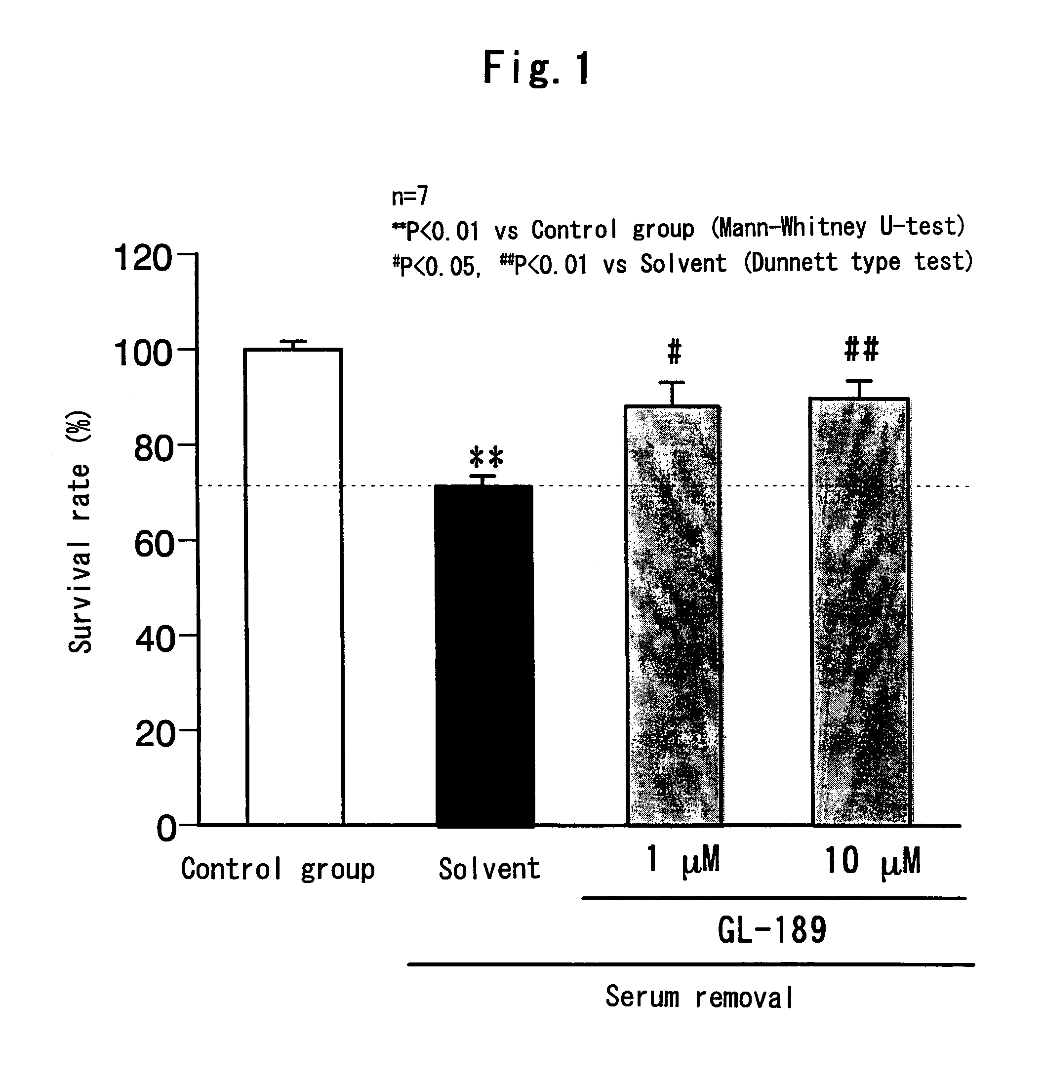
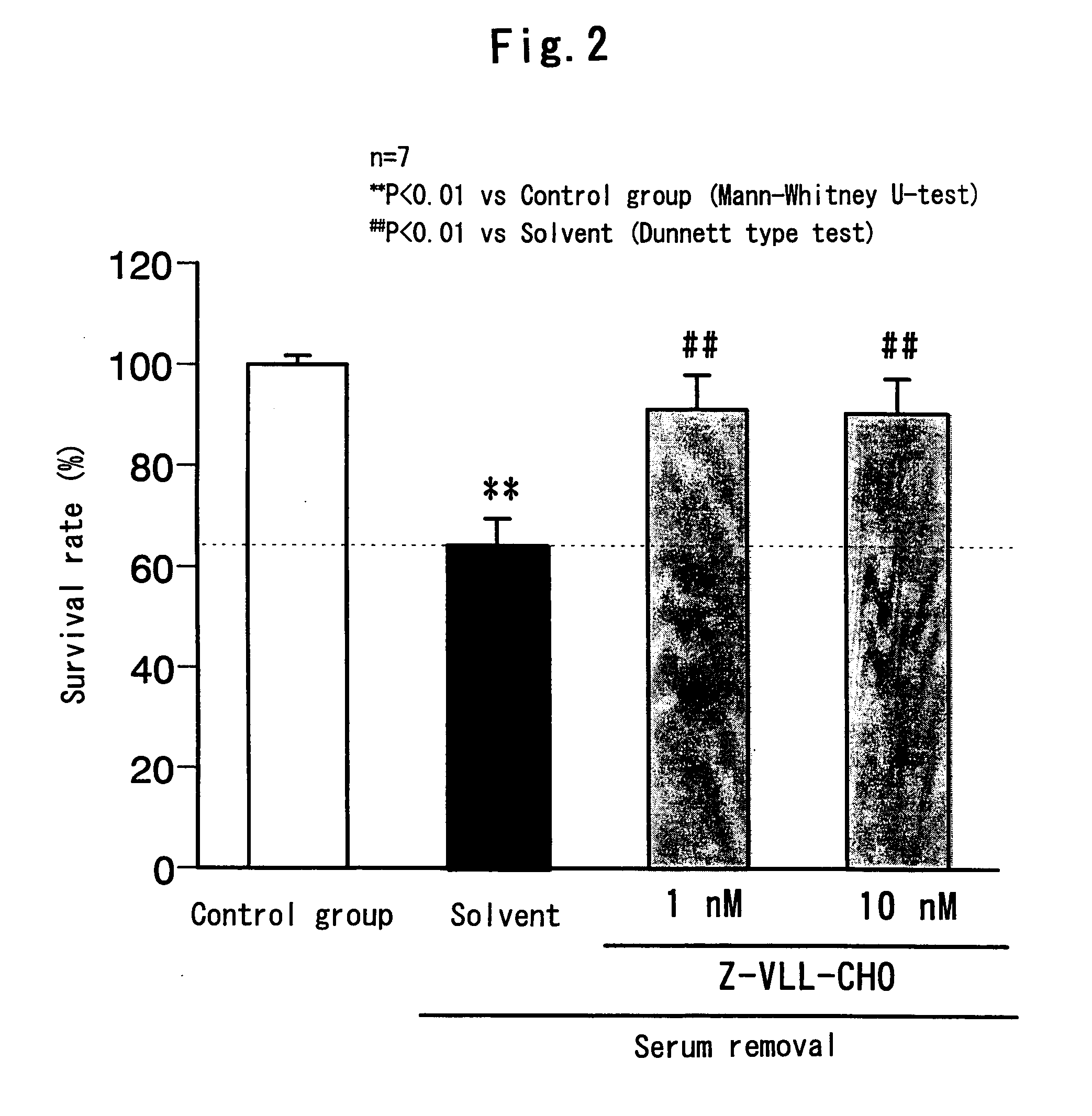
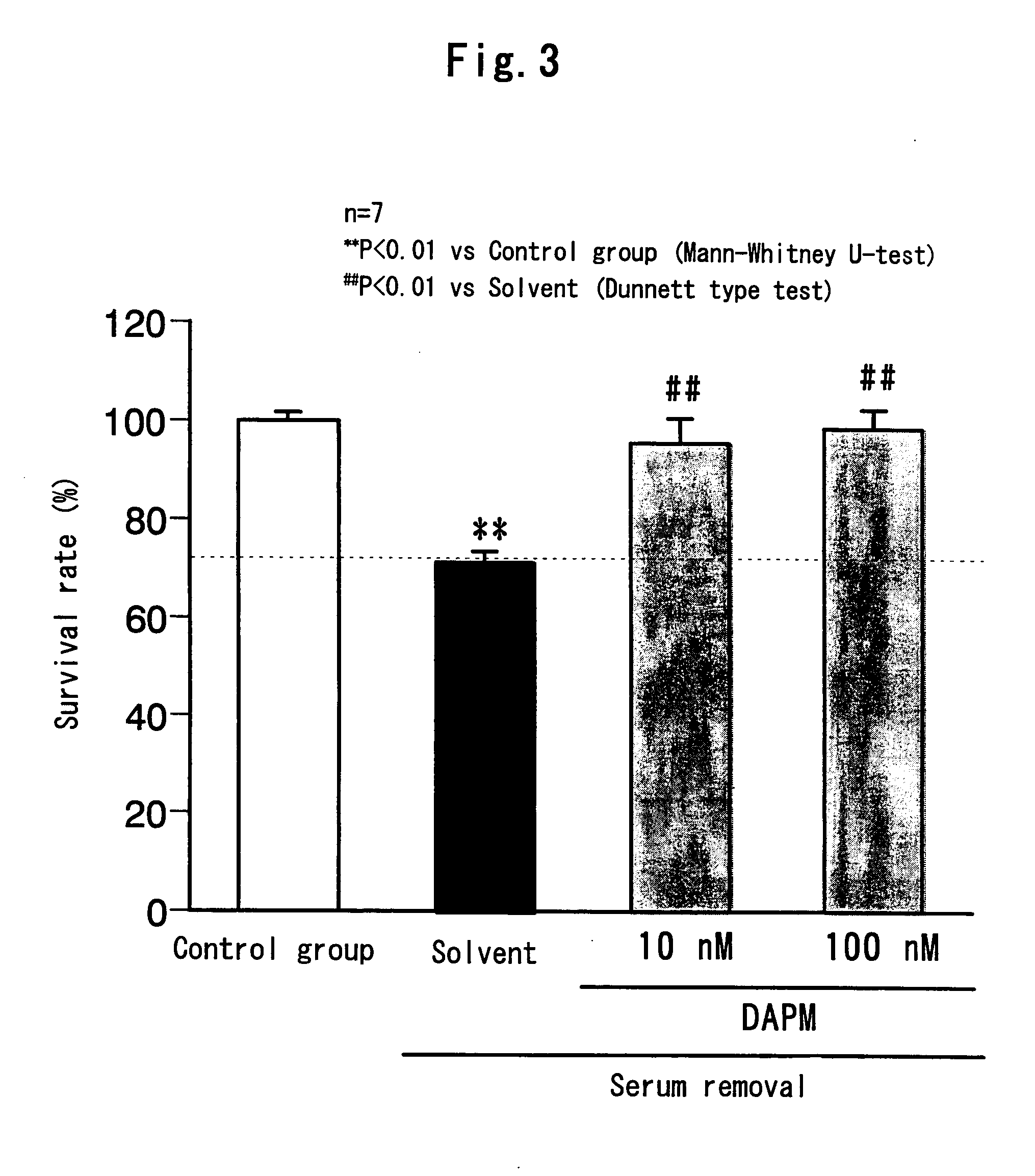
[0035] According to the test method described in Brain Res., 967, 257-66, 2003, retinal neurons of a rat embryo (at 18 days of age) were isolated and seeded on a plastic cover slip coated with polyethylenimine. Then, the cells were cultured for 5 days in an Eagle's minimum essential medium containing 10% fetal bovine serum. From day 6 of the culture, the cells were cultured in an Eagle's minimum essential medium containing 10% fetal bovine serum supplemented with a cytosine arabinoside (at a concentration of 10 μM in the medium), whereby the growth of non-neuronal cells was inhibited. On day 8 of the culture, the medium was changed to an Eagle's minimum essential medium containing 10% horse serum and cultivation was carried out for 24 hours, and then the cells were used for the test. Incidentally, the cultivation was carried out under the conditions of 37° C. and 5% CO2. On day 9 of the culture, the retinal neurons were incubated (at 37° C. and 5% CO2) for 24 hours in an Eagle's minimum essential medium which contains a solvent (0.1% dimethylsulfoxide) or GL-189 (1 μM and 10 μM) and does not contain serum. Then, the cell viability was determined, and the survival rate of each group to that of the control group (100%) incubated for 24 hours in an Eagle's minimum essential medium containing 10% horse serum supplemented with 0.1% dimethylsulfoxide was calculated. The cell viability was determined by the trypan blue staining method. More specifically, the cells were stained for 10 minutes with 1.5% trypan blue solution, fixed with 10% neutral formalin solution and washed with a physiological saline. A stained cell was determined as a dead cell and an unstained cell was determined as a live cell, and the numbers of dead cells and live cells (the total number of cells was 200 or more) were counted under a microscope. The experimental results are shown in the graph of FIG. 1. The columns in the graph represent the average value for each group, and the error lines represent the standard error.
[0036] The same procedure as that described in (1) above was followed except that Z-VLL-CHO (1 nM and 10 nM) was used instead of GL-189, and the survival rate of each group was calculated. The experimental results are shown in the graph of FIG. 2.
[0037] The same procedure as that described in (1) above was followed except that DAPM (10 nM and 100 nM) was used instead of GL-189, and the survival rate of each group was calculated. The experimental results are shown in the graph of FIG. 3.
[0038] The same procedure as that described in (1) above was followed except that γXIV (1 nM and 10 nM) was used instead of GL-189, and the survival rate of each group was calculated. The experimental results are shown in the graph of FIG. 4.
[0039] According to the test method described in Brain Res., 967, 257-66, 2003, RGCs of a rat neonate (at 7 days of age) were isolated by the two-step panning method. As the culture medium, a neuronal culture medium containing a brain derived neurotrophic factor (BDNF, 50 ng / ml), a ciliary neurotrophic factor (CNTF, 50 ng / ml), L-glutamine (2 mM), penicillin / streptomycin (100 U / ml and 100 μg / ml), forskolin (5 μM) and B-27 supplement was used. After the isolation of RGCs, the cells were incubated for 24 hours in a culture medium containing a solvent (0.1% dimethylsulfoxide) or GL-189 (1 μM). Then, glutamic acid was added to give a final concentration of 25 μM in the culture solution and incubation was further carried out for 2 days. The incubation was carried out under the conditions of 37° C. and 5% CO2. As for the control group, incubation was carried out in a culture medium containing a solvent (0.1% dimethylsulfoxide) for the same period of time. After the glutamic acid treatment, identification of live RGCs was carried out using Calcein-AM. More specifically, the cells were incubated for 15 minutes in 1 μg / ml of Calcein-AM solution and washed with a phosphate buffer saline. A cell which is positive for calcein and has a neurite with a length equal to or greater than the diameter of the cell was considered to be a live RGCs, and the number of live RGCs was counted under a fluorescence microscope, and then the survival rate was calculated by taking the case where the number of live RGCs was the same as in the control group as 100%. The experimental results are shown in the graph of FIG. 5. The columns in the graph represent the average value for each group, and the error lines represent the standard error.
[0040] The same procedure as that described in (1) above was followed except that Z-VLL-CHO (100 nM) was used instead of GL-189, and the survival rate of each group was calculated. The experimental results are shown in the graph of FIG. 6.

Problems solved by technology

Glaucoma is one of the eye diseases which cause serious disturbance of visual function leading to the blindness if it is not treated appropriately.
However, it has not been studied at all that what pharmacological action a compound having a secretase inhibitory activity exhibits on a retinal disease or an eye disease such as glaucoma.

Method used


Image

  • Remedy for eye diseases accompanied by optic nerve injuries
  • Remedy for eye diseases accompanied by optic nerve injuries
  • Remedy for eye diseases accompanied by optic nerve injuries

Examples


preparation examples

Preparation Example 1

Eye Drop

[0044]

Formulation:GL-189 1 mgConcentrated glycerin250 mgPolysorbate 80200 mgSodium dihydrogenphosphate 20 mgdihydrate1 N sodium hydroxideq.s.1 N hydrochloric acidq.s.Sterile purified waterq.s.Total 10 ml

[0045] To sterile purified water, GL-189 and the above-mentioned components other than GL-189 were added and mixed thoroughly to prepare an eye drop.

preparation example 2

Tablet

[0046]

Formulation:DAPM1mgLactose66.4mgCorn starch20mgCarboxymethylcellulose6mgcalciumHydroxypropylcellulose6mgMagnesium stearate0.6mgTotal100mg

[0047] DAPM, lactose and corn starch were mixed in a mixer, and then carboxymethylcellulose calcium and hydroxypropylcellulose were added to the mixture and granulated. The resulting granules were dried and sized, and then magnesium stearate was added to the sized granules followed by mixing. Then, the mixture was compressed into a tablet with a tableting machine.


PUM

PropertyMeasurementUnit
thicknessaaaaaaaaaa
flow rateaaaaaaaaaa
flow rateaaaaaaaaaa

Abstract

An object of the present invention is to search for a novel medicinal use of a compound having a secretase inhibitory activity. Because the compound having a secretase inhibitory activity has a remarkable inhibitory effect on retinal neuronal cell death, it is useful as a therapeutic agent for a retinal disease typified by retinal vessel occlusion, diabetic retinopathy, ischemic optic neuropathy, macular degeneration, retinitis pigmentosa and Leber's disease or an eye disease accompanied by an optic neuropathy such as glaucoma.

Description

TECHNICAL FIELD [0001] The present invention relates to a protective agent for retinal neurons comprising a secretase inhibitor as an active ingredient and in particular to a therapeutic agent for an eye disease accompanied by an optic neuropathy such as a retinal disease or glaucoma. BACKGROUND ART [0002] The retina has the function of receiving light from outside and plays an important role for visual function. Structurally, it is a tissue with a thickness of 0.1 mm to 0.5 mm consisting of 10 layers including a pigmented epithelial layer of retina, an inner plexiform layer, a retinal ganglion cell layer, a retinal nerve fiber layer and others. The inner plexiform layer has neurons, which are called an amacrine cell and form a synapse by forming a pair with a ganglion cell projection. Because the neurons are highly responsive at the time of the initiation and termination of light illumination, they are thought to function as a detector of light intensity. The retinal ganglion cell ...

Claims


Application Information

Patent Timeline
Patent Type & Authority Applications(United States)
IPC IPC(8): A61K38/08A61K38/06A61K38/55A61P9/10A61P25/02A61P27/02A61P27/06A61P43/00
CPCA61K38/55A61K38/07A61K38/06A61P9/10A61P25/02A61P27/02A61P27/06A61P43/00
Inventor YONEDA, SHINJIYAMAMOTO, RIEHARA, HIDEAKI
Owner SANTEN PHARMA CO LTD
Why Eureka
Try Eureka