Self-closing surgical clip for tissue

a tissue and surgical technology, applied in the field of tissue sealing devices and methods, can solve the problems of difficulty in performing arterial replacement or bypass grafting, difficulty in constructing an arterial anastomosis, and almost a technical impossibility using minimally invasive techniques, so as to facilitate interrupted anastomosis, promote intima-to-intima contact, and reduce the contact between intraluminal metallic components

Inactive Publication Date: 2006-12-28
MEDTRONIC INC
View PDF99 Cites 98 Cited by
  • Summary
  • Abstract
  • Description
  • Claims
  • Application Information

AI Technical Summary

Benefits of technology

[0014] According to yet another aspect of the invention, a fastener is provided that can be delivered to a wound site and can be used to clip tissues to promote intima-to-intima contact. Another aspect is the providing of a fastener that reduces intraluminal metallic component contact.
[0015] Yet another aspect of the present invention is to provide clips that can be used to perform anastomosis with fewer clips that other fastener systems.
[0016] According to another aspect of the invention, a tissue approximation device is provided that facilitates interrupted anastomosis without know tying and which promotes the dilation and growth of the vessel. Another aspect of the invention supplies a self-closing fastener to tissue that securely anchors the fastener to the tissue with a controlled approximation force.

Problems solved by technology

Constructing an arterial anastomosis is technically challenging for a surgeon in open surgical procedures, and is almost a technical impossibility using minimally invasive techniques.
Many factors contribute to the difficulty of performing arterial replacement or bypass grafting.
If one of the tissues is affixed too close to its edge, the suture can rip through the tissue and impair both the tissue and the anastomosis.
Another factor is that, even after the tissues are properly aligned, it is difficult and time consuming to pass the needle through the tissues, form the knot in the suture material, and ensure that the suture material does not become tangled.
These difficulties are exacerbated by the small size of the artery and graft.
Another factor contributing to the difficulty of such procedures is the limited time available to complete the procedure.
The time the surgeon has to complete an arterial replacement or bypass graft is limited because there is no blood flowing through the artery while the procedure is being done.
If blood flow is not promptly restored, sometimes in as little as thirty minutes, the tissue the artery supplies may experience significant damage, or even death (tissue necrosis).
In addition, arterial replacement or bypass grafting is made more difficult by the need to accurately place and space many sutures to achieve a permanent hemostatic seal.
A drawback to this procedure is the difficulty in preventing the suture from tangling and the time and surgical skill required to tie individual knots when using multiple sutures.
Due to space requirements, this procedure is generally limited to open surgery techniques.
The difficulty of suturing a graft to an artery using minimally invasive surgical techniques has effectively prevented the safe use of this technology in both peripheral vascular and cardiovascular surgical procedures.
When manipulating instruments through cannulas, it is extremely difficult to position tissues in their proper alignment with respect to each other, pass a needle through the tissues, form a knot in the suture material once the tissues are aligned, and prevent the suture material from becoming tangled.
Therefore, although there have been isolated reports of vascular anastomoses being formed by minimally invasive surgery, no system has been provided for wide-spread surgical use which would allow such procedures to be performed safely within the prescribed time limits.
It is extremely difficult to perform such positioning techniques in minimally invasive procedures.

Method used


Image

  • Self-closing surgical clip for tissue
  • Self-closing surgical clip for tissue
  • Self-closing surgical clip for tissue

Examples


Embodiment Construction

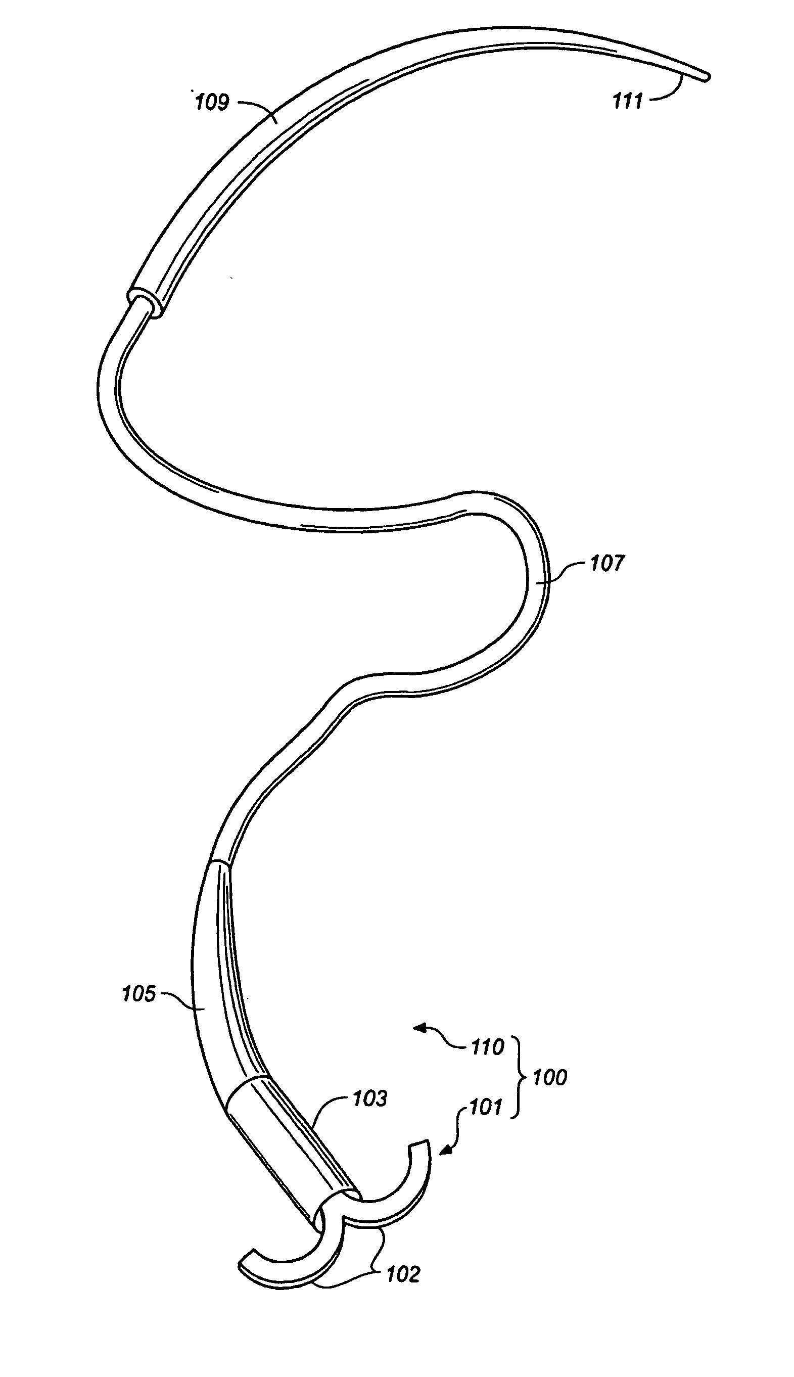
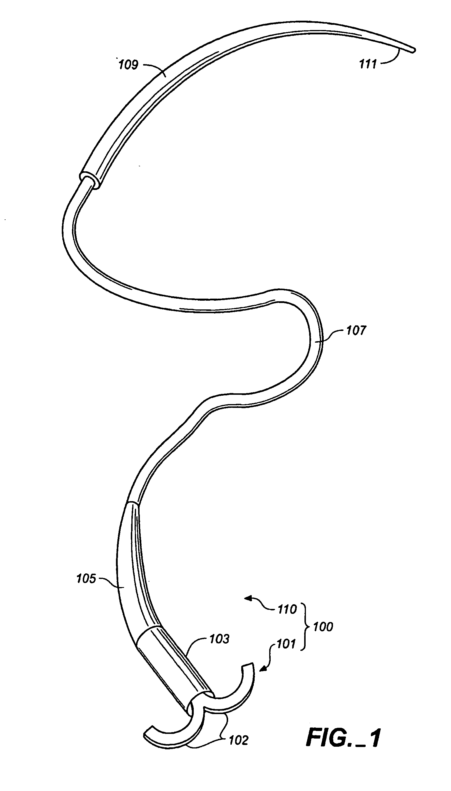
[0042] Referring now to the drawings, and first to FIG. 1, a tissue connector assembly constructed according to the principles of the present invention is shown and generally indicated with reference numeral 100. The tissue connector assembly 100 may be used to manipulate and align tissues, or tissue and graft with respect to each other and thereafter connect the tissues together (FIGS. 6-8). As used herein, the term graft includes any of the following: homografts, xenografts, allografts, alloplastic materials, and combinations of the foregoing. The tissue connector assembly and connectors of the present invention are generally useful for attaching tissues and, as will become apparent upon reflection of the present disclosure, can be used for a variety of medical procedures or can be modified within the scope of the present invention to perform such procedures. Thus the tissue connector assembly 100 may be used as illustrated in FIGS. 7, 8 and 9 in vascular surgery to replace or byp...


PUM


Abstract

A self-closing fastener is described that comprises a clip passable through a tissue opening. The fastener is adapted for holding by a mechanism in an open configuration for passing through the tissue, followed by releasing the fastener from the holding mechanism, allowing the clip to remain in the tissue in a shape that can clip two or more locations on the tissue. The fastener and delivery devices are particularly useful for tissue approximation, such as anastomosis. When used for anastomosis, the inventive clips provide intima-to-intima contact with a minimal amount of intraluminal exposure.

Description

FIELD OF THE INVENTION [0001] The present invention relates to devices and methods for sealing tissue punctures. More specifically, the present invention is directed to devices and methods for approximating wound edges of vessel openings to affect hemostasis. BACKGROUND OF THE INVENTION [0002] Minimally invasive surgery has allowed physicians to carry out many surgical procedures with less pain and disability than conventional, open surgery. In performing minimally invasive surgery, the surgeon makes a number of small incisions through the body wall to obtain access to the tissues requiring treatment. Typically, a trocar, which is a pointed, piercing device, is delivered into the body with a cannula. After the trocar pierces the abdominal or thoracic wall, it is removed and the cannula is left with one end in the body cavity, where the operation is to take place, and the other end opening to the outside. A cannula has a small inside diameter, typically 5-10 millimeters, and sometime...

Claims


Application Information

Patent Timeline
Patent Type & Authority Applications(United States)
IPC IPC(8): A61B17/08
CPCA61B17/0469A61B17/064A61B17/11A61B2017/1135A61B17/1285A61B2017/1107A61B17/1227
Inventor AINSWORTH, STEPHENYANG, JIANHUA
Owner MEDTRONIC INC
Why Eureka
Try Eureka