Biological artificial ligament and method of making

a biochemical and artificial ligament technology, applied in the field of artificial ligaments, can solve the problems of ligament erosion, high incidence of tear of anterior cruciate ligaments located at the knee joints, and further structural damag

Active Publication Date: 2007-02-01
SUMMIT GD BIOTECH
View PDF32 Cites 38 Cited by
  • Summary
  • Abstract
  • Description
  • Claims
  • Application Information

AI Technical Summary

Benefits of technology

[0012] In order to accomplish the objects of the present invention, the present invention provides a biological prosthesis for implantation into a human body which is made by a method that includes the steps of providing a natural animal ligament or tendon that has a substrate, crosslinking and fixing the substrate, minimizing the antigens from the substrate, and coupling an active layer to the substrate.

Problems solved by technology

A torn ligament is one of the most common sport injuries, and the anterior cruciate ligaments located at the knee joints have a very high incidence of tears during sport activities.
The consequences of these tears are usually very severe which can lead to further structural damage such as ligament erosion, impaired joint function, and even disability, if treatment is not performed promptly and properly.
Previously, surgeons attempted treatment by sewing the torn parts, but because ligaments have poor adherence and healing power, almost all of these attempts failed, so that such practices were abandoned.
However, the biggest drawback of such a method is that it increases autoinjuries and leaves complications, sometimes to the extent that the drawbacks outweigh the benefits.
For example, using the patellar tendon as a replacement (in general, the connecting bones are also removed in the form of bone-patellar tendon-bone) for transplantation easily causes complications such as knee pain, patellar tendon contraction, and tendonitis, and complete success cannot be insured if the procedure is not done perfectly.
Another method uses tendons or ligaments as replacements by allogenic transplantation (i.e, from a fresh cadaver), but this method has two important drawbacks: (1) the source is scarce, and (2) the support measures are out-of-date, and very often the specimens are simply stored in low temperature refrigerators and utilized in transplantations after simple sterilization by immersing in a sterilizing solution, which cannot effectively eliminate antigens and kill all the bacteria and viruses which might be present in the cadaver body, nor can it prevent rejection or eliminate the risk of infections after transplantation.
However, these synthetic polymers are different from body tissues with respect to their composition and structure, so that the risk of having aseptic inflammation due to chronic rejection is ever present.
More importantly, polymeric materials have inherent creep deformation in nature and after a certain time of playing the function of ligaments, they are irreversibly elongated and lose the elastic function of ligaments.
Glutaraldehyde fixes proteins by crosslinking them and forming aldehyde acetals, but aldehyde acetal is not very stable and the resistance of the fixed substance to degradation is poor, which results in loss of the inherent mechanical strength due to degradation.
In addition, the degradation slowly releases glutaraldehyde and generates toxic aldehyde radicals, and this is compounded by the problem that there is no antigenic treatment in the process to effectively eliminate the antigenicity, so that failures are often experienced due to chronic rejection when the specimens are transplanted into the body.

Method used


Image

  • Biological artificial ligament and method of making
  • Biological artificial ligament and method of making
  • Biological artificial ligament and method of making

Examples


example 1

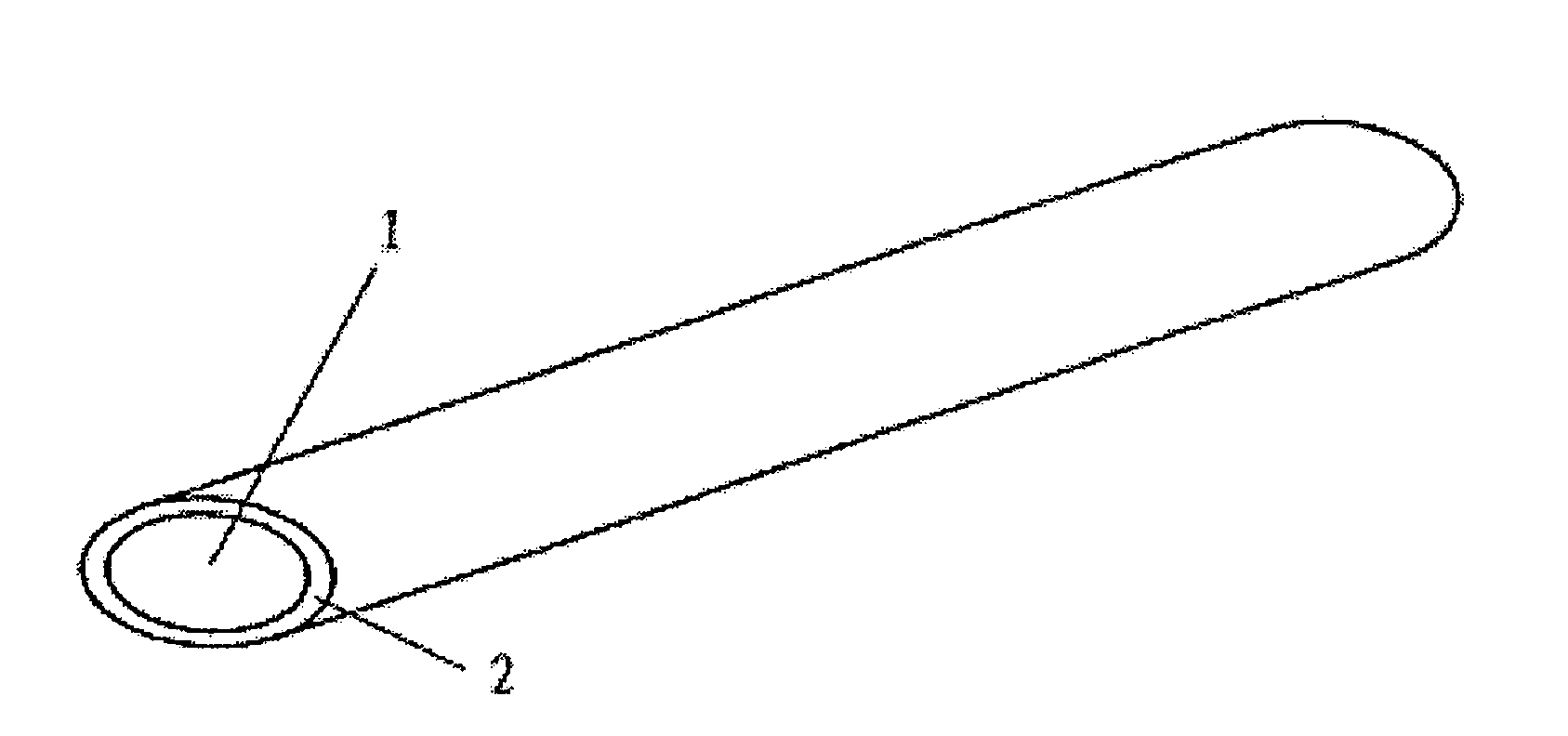
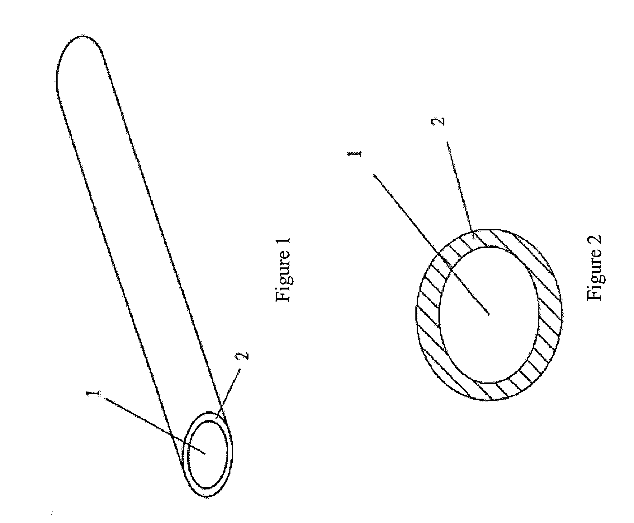
[0043] Referring to FIGS. 1 and 2, a biological artificial ligament comprising a substrate 1 is formed with an animal ligament or tendon crosslinked and fixed with an epoxide and treated to minimize antigens. An activated surface layer 2 is formed on the surface of the substrate 1 by coupling a specific polypeptide capable of adhering to and accumulating growth factors. In this example, the polypeptide is the polypeptide consisting of 16 lysines (K16), glycine (G), arginine (R), aspartic acid (D), serine (S), proline (P) and cysteine (C), and said glucosaminoglycan is hyaluronic acid, chondroitin sulfate, dermatan sulfate, heparin, acetylated heparin sulfate or keratin sulfate. This biological artificial ligament can be made from the following steps:

[0044] 1. Screening of materials: Fresh animal ligaments and tendon are collected by professional technicians from regulated and well-managed slaughterhouses while special efforts are made to avoid direct contact with pollutants.

[0045]...


PUM

PropertyMeasurementUnit
temperatureaaaaaaaaaa
water-solubleaaaaaaaaaa
hydrogen bonding poweraaaaaaaaaa

Abstract

A prosthesis for implantation into a human body is made by a method that includes the steps of providing a natural animal ligament or tendon that has a substrate, crosslinking and fixing the substrate, minimizing the antigens from the substrate, tanning the substrate to improve its mechanical properties, and coupling an active layer to the substrate.

Description

BACKGROUND OF THE INVENTION [0001] 1. Field of the Invention [0002] The present invention relates to a medical prosthesis for human implantation, and in particular, to an artificial ligament which is utilized to rebuild the ligament of a patient with a torn ligament. [0003] 2. Description of the Prior Art [0004] A torn ligament is one of the most common sport injuries, and the anterior cruciate ligaments located at the knee joints have a very high incidence of tears during sport activities. The consequences of these tears are usually very severe which can lead to further structural damage such as ligament erosion, impaired joint function, and even disability, if treatment is not performed promptly and properly. Previously, surgeons attempted treatment by sewing the torn parts, but because ligaments have poor adherence and healing power, almost all of these attempts failed, so that such practices were abandoned. [0005] Therefore, surgeons placed their hopes on rebuilding ligaments by...

Claims


Application Information

Patent Timeline
Patent Type & Authority Applications(United States)
IPC IPC(8): A61F2/08
CPCA61F2/062A61F2/08A61L27/227Y10S623/915A61L27/3683A61L2430/10A61L27/3604A61P43/00
Inventor XU, GUO-FENG
Owner SUMMIT GD BIOTECH
Why Eureka
Try Eureka