Inducing expression of puma to reduce joint inflammation in the treatment of arthritis
a technology of puma and expression, applied in the field of arthritis treatment, to achieve the effect of reducing the pro-inflammatory and destructive activity
- Summary
- Abstract
- Description
- Claims
- Application Information
AI Technical Summary
Benefits of technology
Problems solved by technology
Method used
Image
Examples
example 1
Materials and methods
[0090] Synovial tissue samples were obtained from patients with osteoarthritis (OA) and rheumatoid arthritis (RA) at the time of joint replacement. The diagnosis of RA conformed to the 1987 revised American College of Rheumatology criteria (Arnett et al., Arthritis Rheum (1988) 31:315-324). Normal human spleen tissue was provided by the UCSD Cancer Center, San Diego tissue bank. The studies were approved by the University of California, San Diego, Human Subjects Research Protection Program. FLS used for experiments were prepared from enzymatically dispersed synovial tissue by treating the tissues with 1 mg / ml collagenase and culturing in DMEM supplemented with 10% fetal calf serum (FCS), penicillin, streptomycin, and L-glutamate. Cell lines were used from the 3rd through 9th passage, when they are a homogenous population of fibroblast like cells. Normal Human skin fibroblasts were purchased from Cell Applications (San Diego, Calif.). p53+ / +and p53− / −murine syno...
example 2
PUMA Regulation and Proapoptotic Effects in Fibroblast-like Synoviocytes
[0098] In this example, experiments were performed to assess PUMA expression in synovial tissue obtained from patients with rheumatoid arthritis.
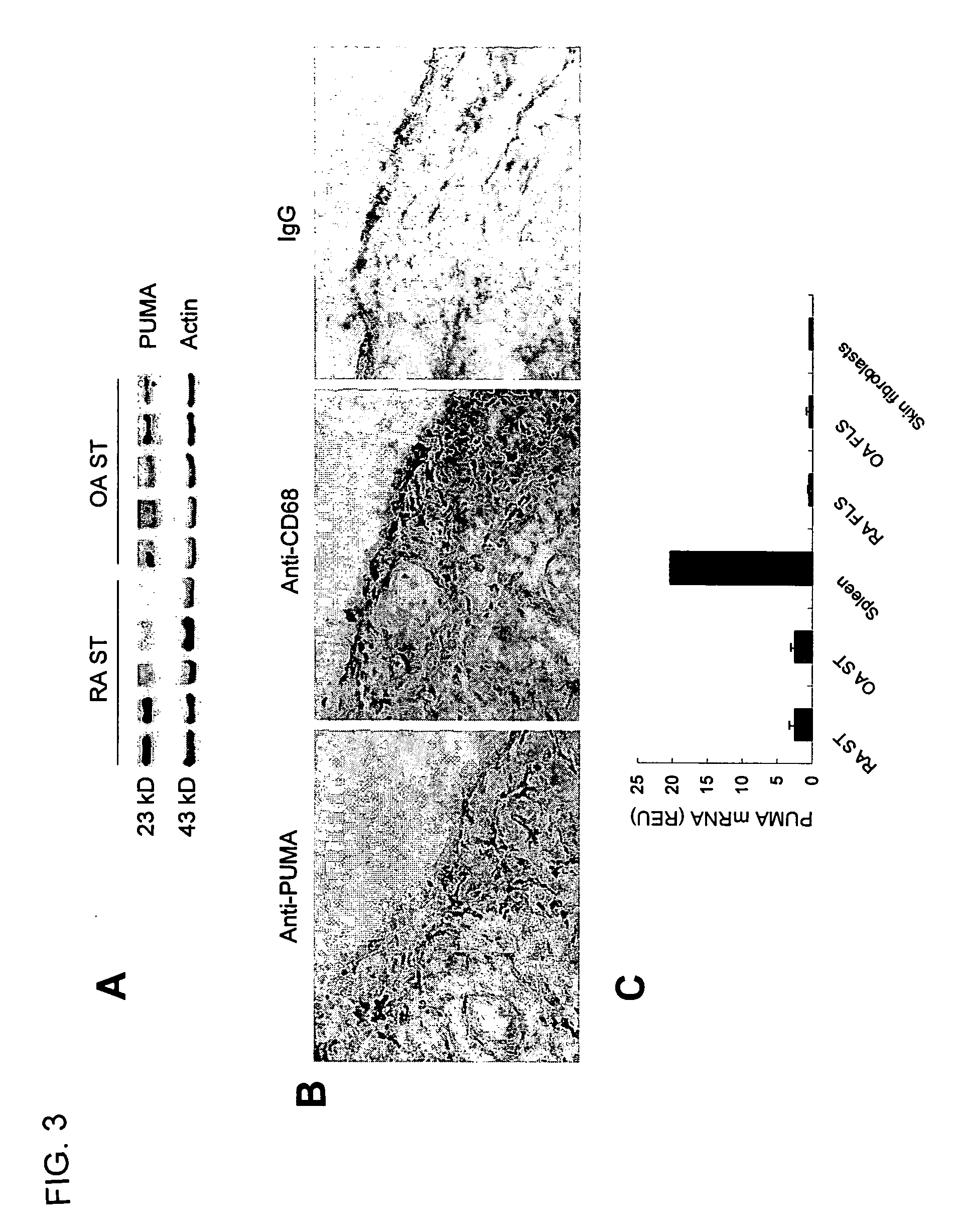
[0099]FIG. 3 shows PUMA protein and gene expression in RA synovium and FLS. FIG. 3(A): Total protein from RA (n=5) and OA (n=5) ST was extracted and Western blot analysis was performed to detect PUMA and β-actin expression. FIG. 3(B): A representative example of immunohistochemistry to detect PUMA is shown for RA ST. Although expression was low, when PUMA was detected in RA synovium it was mainly expressed the synovial sublining area. For comparison to demonstrate the intimal lining in the same tissue, CD68 expression in the lining cells is also shown. A serial section with an isotype matched control antibody (IgG) was negative. FIG. 3(C): Total RNA was isolated from RA ST (n=16), OA ST (n=15), normal human spleen tissue, RA FLS (n=5), OA FLS (n=5), and normal skin fi...
example 3
PUMA Regulation and Proapoptotic Effects in fibroblast-like Synoviocytes
[0113] Experiments were performed to determine whether siRNA could knock down p53 expression in cultured FLS.
[0114]FIG. 6 shows a representative time course of the effect of siRNA p53 knockdown on cultured FLS. FIG. 6(A): Western blot analysis. Cultured FLS were transfected with 1, 2.5, or 5 μg of siRNA or non-silence scrambled siRNA (sc). Mock transfected cells were treated in the same manner except that no siRNA was added. FLS were then incubated for 3 or 5 days and Western blot analysis was performed. FIG. 6(B): Irnmunohistochemistry staining of p53 protein expression in RA FLS. Transfected FLS were seeded into 4-well chamber slides, cultured for 5 days, and evaluated by immunohistochemistry. Manse percentage of p53-positive cells is shown.
[0115] The residual expression of p53 protein was approximately 8-10%, 5%, and 1-2% for 1, 2.5, and 5 μg of siRNA as determined by Western blot analysis. Immunohistochem...
PUM
| Property | Measurement | Unit |
|---|---|---|
| Composition | aaaaa | aaaaa |
Abstract
Description
Claims
Application Information
 - R&D
- Intellectual Property
- Life Sciences
- Materials
- Tech Scout
- Unparalleled Data Quality
- Higher Quality Content
- 60% Fewer Hallucinations
Browse by: Latest US Patents, China's latest patents, Technical Efficacy Thesaurus, Application Domain, Technology Topic, Popular Technical Reports.
© 2025 PatSnap. All rights reserved.Legal|Privacy policy|Modern Slavery Act Transparency Statement|Sitemap|About US| Contact US: help@patsnap.com


